. 2020 Nov 2;9:e56059. doi: 10.7554/eLife.56059

Figure 4. Expression of corin mutants with point mutations in the DSSDE motif in LDLR8 module.

(A) Corin wild-type (WT) and mutants with point mutations at indicated positions (D682A, S683A, S684A, D685A, and E686A), indicated in red, were expressed in MDCK cells. Corin expression (green) on apical and basolateral membranes was examined by immunostaining and confocal microscopy with X-Y (top panels) and X-Z (lower panels) views. (B) Corin WT and S684A mutants without (S684A) or with the DSSDE motif created in LDLR6 (ML6/S684A) or LDLR7 (ML7/S684A) module expressed in MDCK cells were examined by immunostaining and confocal microscopy. X-Y (top panels) and X-Z (lower panels) views are indicated. Each image represents the data from five experiments. Scale bars: 5 μm.

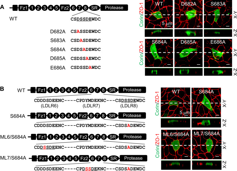
Figure 4.

Figure 4—figure supplement 1. Analysis of the DSSDE motif in corin LDLR8 module.

Figure 4—figure supplement 1.

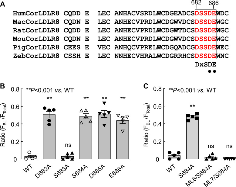
(A) Alignment of the DSSDE motif in corin LDLR8 modules from selected vertebrate species. The DSSDE motif (red) in corin LDLR8 modules from selected vertebrate species (human, Hum; monkey, Mac; rat, Rat; mouse, Mou; pigeon, Pig; zebrafish, Zeb) is aligned. This motif corresponds to the DxSDE motif found in LDLR modules in many proteins. The Asp-Glu residues (indicated by black dots), corresponding to Asp685 and Glu686 in human corin, have been shown to be part of a Ca2+-binding cage in LDLR module structures of other proteins. (B and C) Corin WT and mutants were expressed in MDCK cells and analyzed by immunostaining and confocal microscopy. Quantitative data of relative corin expression on apical and basolateral membranes, indicated by the ratio of FBL/FTotal, were analyzed by Image J software. The data (mean ± SEM) were from five experiments. Statistical analysis was done with ANOVA. n.s., not significant vs. corin WT in respective groups.
Figure 4—figure supplement 1—source data 1. Source data for Figure 4—figure supplement 1B.
Figure 4—figure supplement 1—source data 2. Source data for Figure 4—figure supplement 1C.
Figure 4—figure supplement 2. Western blotting of corin proteins in biotin-labeled cell membranes.

Figure 4—figure supplement 2.

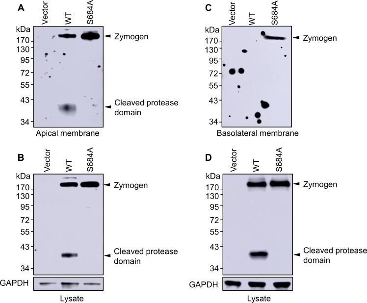
MDCK cells stably transfected with a vector or plasmids expressing corin WT and the S684A mutant in Transwell plates were treated with sulfo-NHS-biotin in upper (A and B) or lower (C and D) chambers to label apical and basolateral membrane proteins, respectively. The cells were lysed. Biotin-labeled corin proteins (A and C) were immunoprecipitated and analyzed by western blotting. In parallel, corin proteins in cell lysates were analyzed by western blotting with GAPDH control (B and D). The data are representative of three experiments.