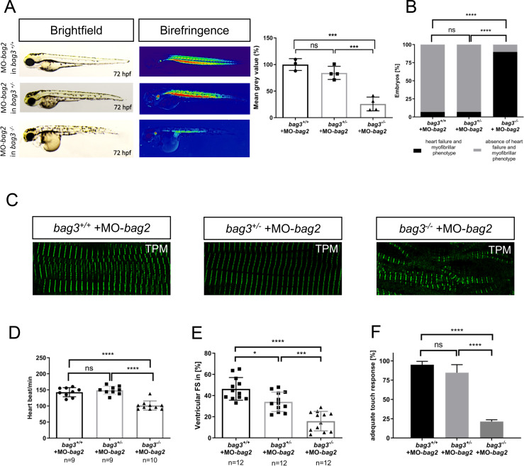
Fig 7. Knockdown of bag2 in bag3-/- mutants causes heart and skeletal muscle disruptions.
(A) Brightfield and birefringence images and densitometric analysis of birefringence (n = 4) of bag3+/+, bag3+/- and bag3-/- embryos at 72 hpf injected with 200μM of MO-bag2. Representative samples are shown (One-way ANOVA followed by tukey's multiple comparison analysis, P<0.0004). (B) bag3-/- embryos injected with MO-bag2 develop (cardio-)myopathy (89.76±4.37%), whereas bag3+/+ (93.10±0.33%) and bag3+/- (94.77±2.35%) embryos are unaffected by MO-bag2 injection (N = 3, n = 50, mean ± S.D., P<0.0001, two-tailed value for Fisher´s exact test). (C) Tropomyosin immunostainings of bag3+/+, bag3+/- and bag3-/- embryos injected with MO-bag2 at 72 hpf. Only MO-bag2-injected bag3-/- embryos show muscle fiber disruptions. (D) Heart rate quantification at 72 hpf reveals impairments only in bag3-/- embryos injected with MO-bag2 (N = 3, n = 9/10, mean ± S.D. One-way ANOVA followed by tukey's multiple comparison analysis, P<0.0001). (E) FS of ventricles of bag2 splice MO injected in bag3-/- embryos at 72 hpf (FS: 15.83±9.19%) is significantly reduced compared to the FS measured in bag3+/+ (FS: 46.33±10.72%) and bag3+/- (FS: 34.07±9.17%) injected embryos (N = 3, n = 12; mean± SD One-way ANOVA followed by tukey's multiple comparison analysis, *P = 0.0110,***P = 0.0002, ****P< 0.0001). (F) bag3-/- embryos injected with MO-bag2 reveal significantly impaired responsiveness upon mechanical stimulus compared to bag3+/+and bag3+/- embryos injected with MO-bag2 (N = 3, n = 80/50, mean ± S.D. One-way ANOVA followed by tukey's multiple comparison analysis, P<0.0001).

