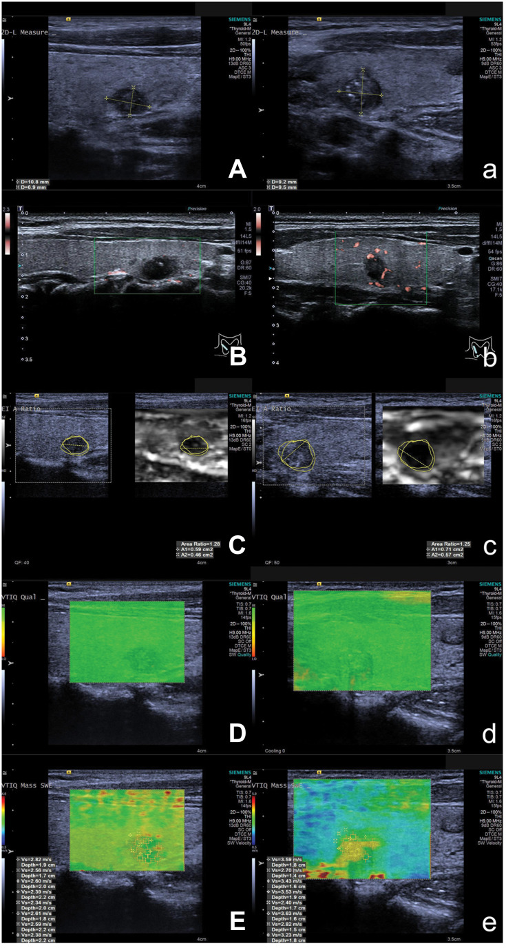
Figure 2.
Ultrasound and ARFI elastography images of patients with solitary and cN0 PTC. Figures (A–E) are images of contralateral CLMN in patients with solitary and cN0 PTC, while Figures (a) to (e) are images without contralateral CLMN. Grayscale ultraound, SMI, VAR displayed on VTI, ARFI quality mode, VTIQ are showed in figures (A) and (a), (B) and (b), (C) and (c), (D) and (d), and (E) and (e). The images of grayscale ultrasound, SMI, and VAR are similar between patients with or without contralateral CLMN. However, the SWVmean of patient with contralateral CLMN (3.17 m/s) displayed on VTIQ is higher than that of patient without contralateral CLMN (2.54 m/s).
Abbreviations: ARFI, acoustic radiation force impulse; cN0, clinical lymph node negative; PTC, papillary thyroid carcinoma; CLMN, central lymph node metastasis; SMI, superb microvascular imaging; VTI, virtual touch tissue; VTIQ, virtual touch tissue quantification; VAR, virtual touch tissue area ratio; SWV, shear wave velocity.

