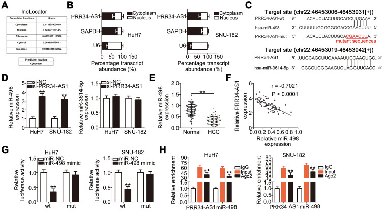
Figure 2.
PRR34-AS1 functions as an miR-498 sponge in HCC cells. (A) lncLocator predicted the subcellular distribution of PRR34-AS1. (B) Cellular nucleocytoplasmic fractionation was performed to isolate the nuclear and cytoplasmic fractions of HuH7 and SNU-182 cells. Both fractions were analyzed via RT-qPCR to determine the localization of PRR34-AS1in HCC cells. (C) The complementary binding sites of miR-498 and miR-3614-5p within PRR34-AS1. (D) miR-498 and miR-3614-5p expression in PRR34-AS1-depleted HuH7 and SNU-182 cells were measured via RT-qPCR. (E) RT-qPCR analysis was used to measure the expression of miR-498 in 65 pairs of HCC and adjacent normal tissues. (F) The correlation between miR-498 and PRR34-AS1 levels in the 65 HCC tissues was analyzed using Pearson’s correlation coefficient. (G) Luciferase reporter assays were used to analyze the binding interaction between miR-498 and PRR34-AS1 in HCC cells. Cotransfection of miR-498 mimic and the wt-PRR34-AS1 reporter plasmid clearly reduced the luciferase activity in HuH7 and SNU-182 cells. The luciferase activity of the mut-PRR34-AS1 reporter plasmid was unchanged after miR-498 mimic cotransfection. (H) RIP assay was conducted using the anti-Ago2 antibody to determine the enrichment of miR-498 and PRR34-AS1 in HuH7 and SNU-182 cells. **P < 0.01.

