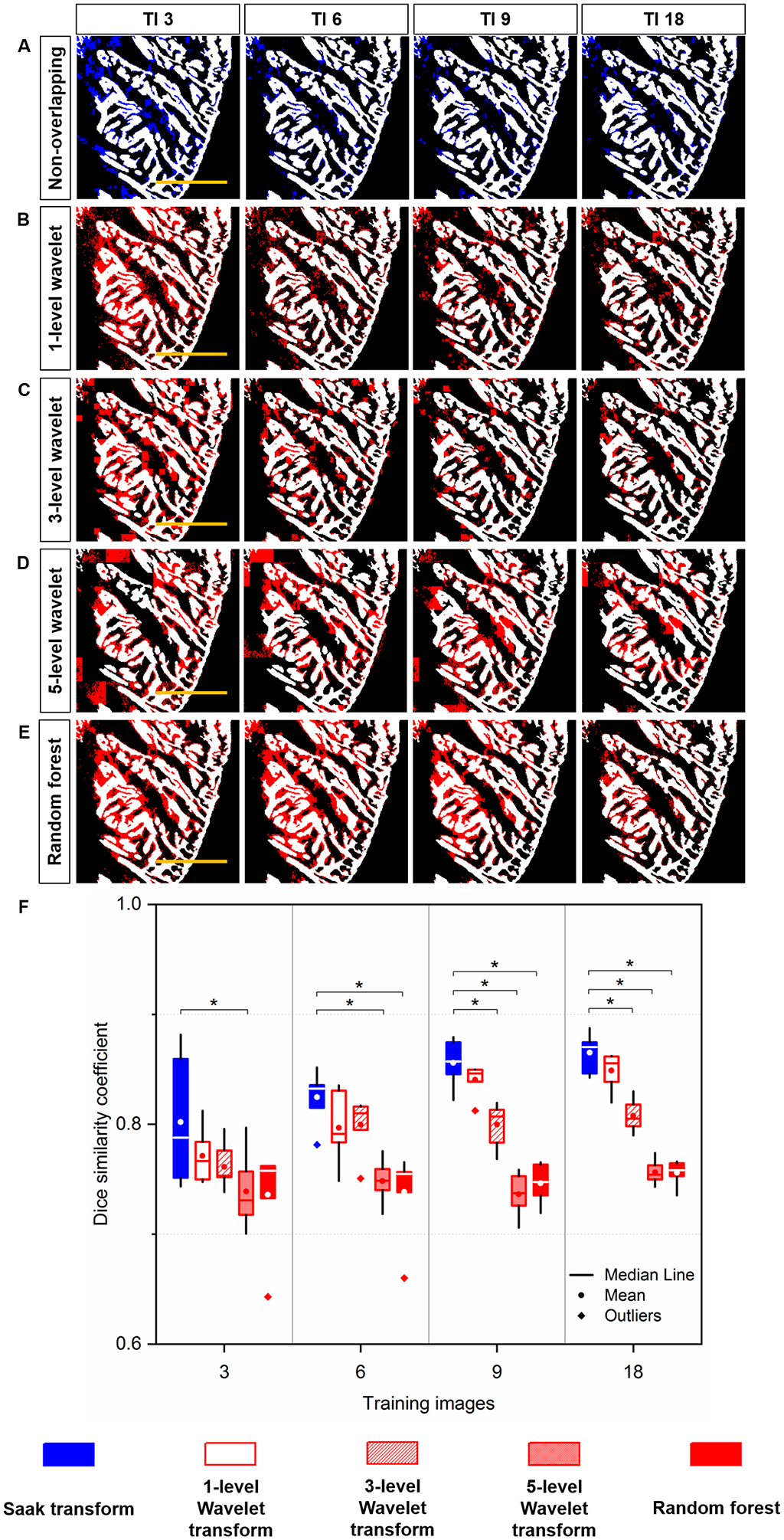
Fig 5.

Quantitative comparison of Segmentation: (A) Saak transform (in blue) vs. (B-D) Wavelet transform (in red) and (E) Random forest (in red) following 3, 6, 9 and 18 training images. These segmentation results were merged with the manually annotated image (in white) for quantitative comparison. (F) Saak transform-based method (random forest in combination with Saak coefficients) outperforms 3- and 5-level Haar Wavelet transforms (random forest in combination with Wavelet coefficients) starting from 9 and 3 training images, respectively. Starting from 6 training images, Saak transform-based method surpasses random forest, indicating the statistically significant difference (p < 0.05, n = 6 testing images for each condition). Scale bar: 200 μm.
