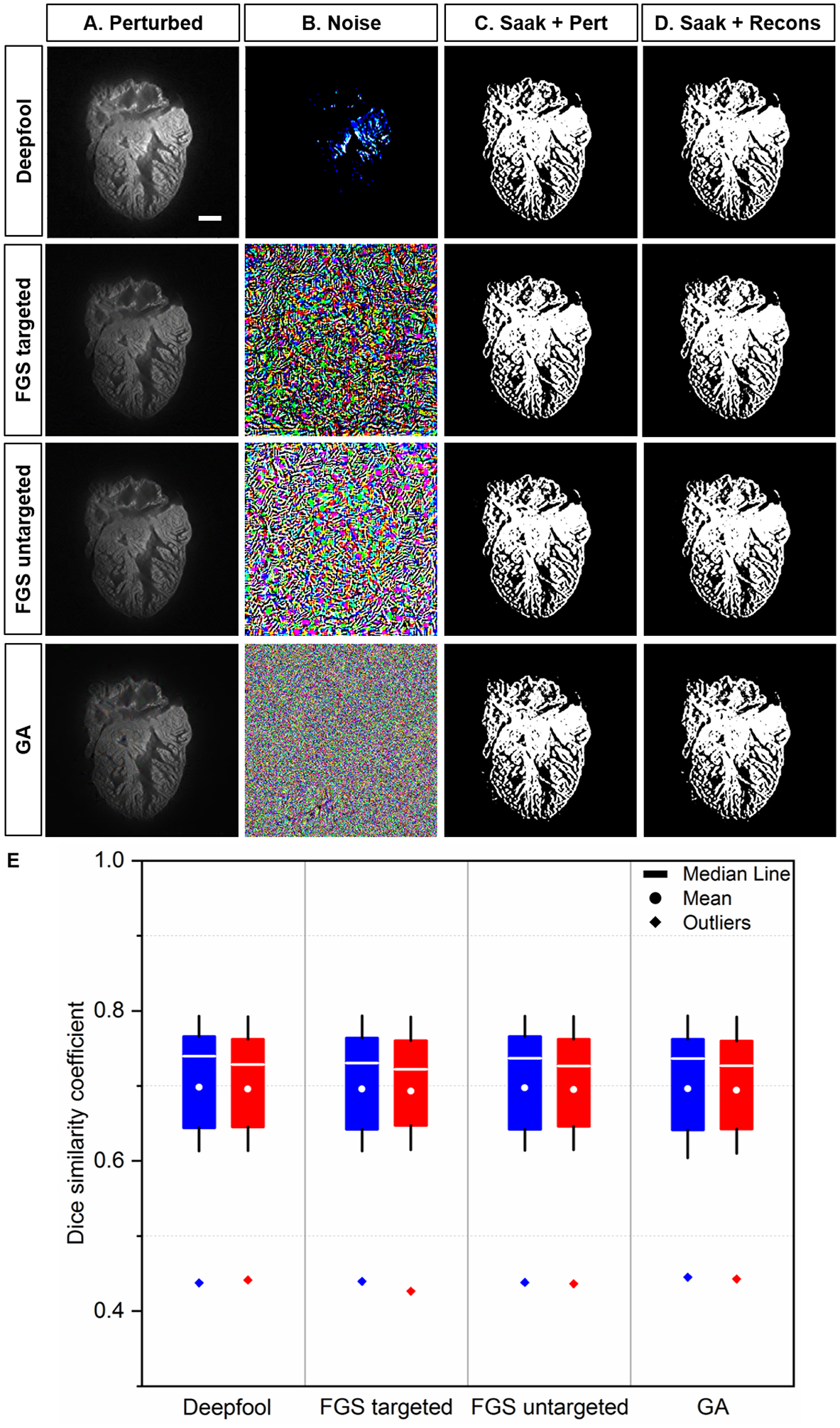
Fig 7.

Saak transform-based segmentation following perturbed and reconstructed images. (A) Perturbed images were generated by introducing the original images to (B) adversarial perturbations from Deepfool, targeted and untargeted fast gradient sign (FGS), and gradient ascent (GA) methods, respectively. (C-D) Non-overlapping Saak transform plus random forest were performed to segment the cardiac trabecular network. (E) The values of DSC between perturbed and reconstructed images were statistically insignificant (p >0.05, n=12 testing images for each perturbation). Blue: perturbed; red: reconstructed. Scale bar: 200 μm.
