
Fig. 6.
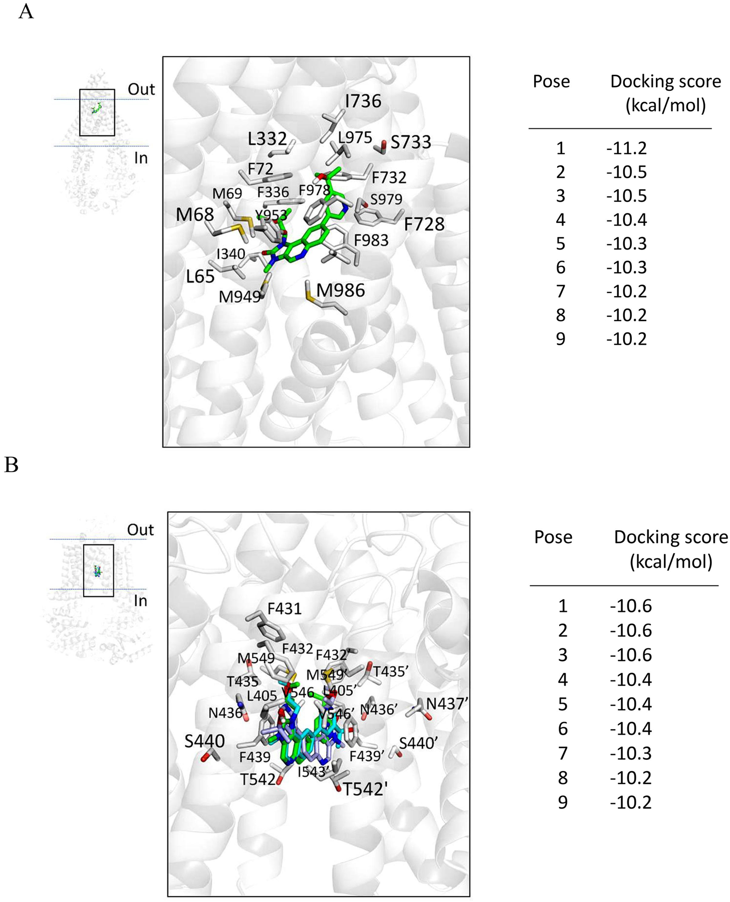
Binding of LY3023414 in the substrate-binding pockets of ABCB1 and ABCG2. LY3023414 was docked to the cryo-electron microscopy inward-open structure of (A) human ABCB1 (PDBID:6QEX) and (B) human ABCG2 (PDB: 5NJ3) using AutoDock Vina software as described in Materials and methods. LY3023414 lowest energy pose binding to ABCB1 is shown in green, whereas the three lowest energy poses of LY3023414 binding to ABCG2, all with the same docking score, are shown in green, cyan, and light blue. Residues that are within 5Å of the ligand are highlighted. The docking scores of the low-energy poses (tighter binding) of LY3023414 in the transmembrane region of ABCB1 and ABCG2 are presented on the right of each panel. Figures were prepared using the PyMOL molecular graphics system, Version 1.7 Shrödinger, LLC.