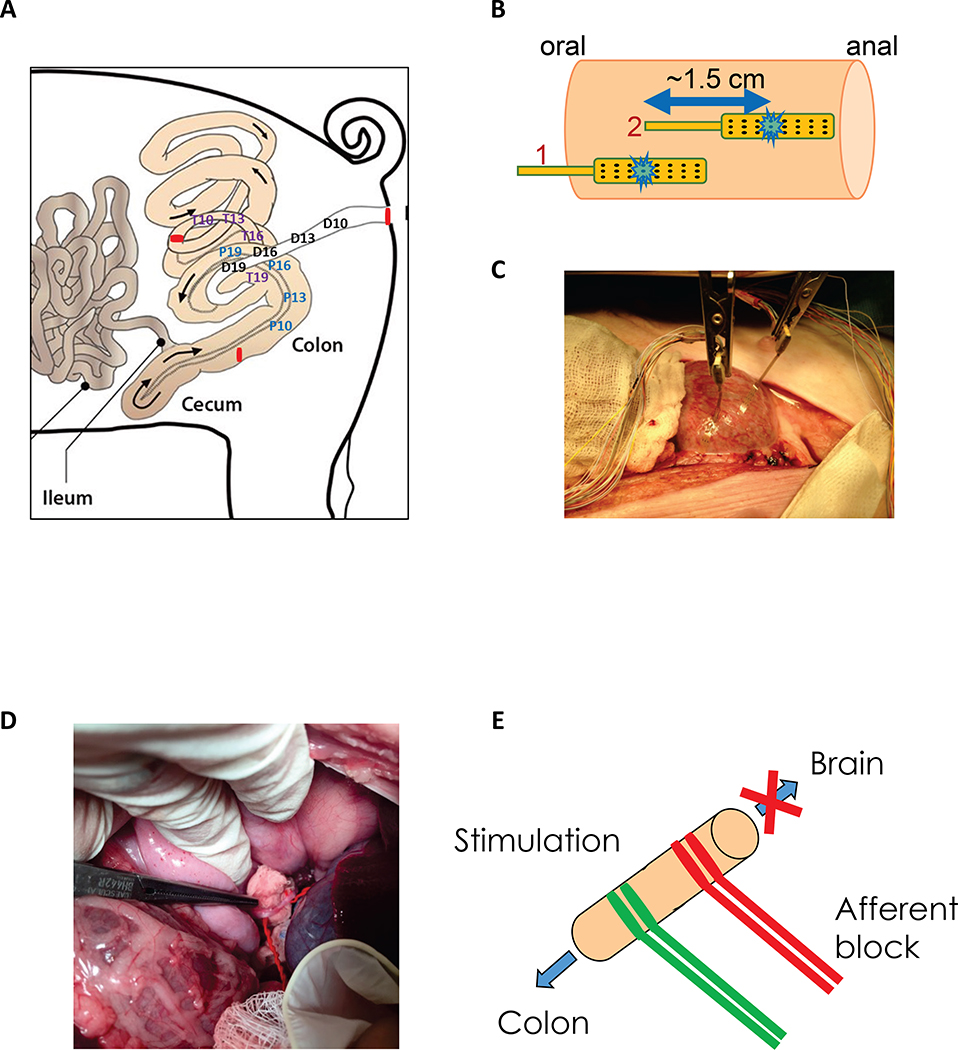
Figure 1. Schematic representation of colonic manometry recordings in overnight fasted and anesthetized adult male castrated Yucatan pigs.
A) Solid state manometry sensors were positioned at 10, 13, 16 and 19 cm from the ceco-colic junction (P10, P13, P16, P19), from the distal end of the proximal/ascending colon (T10, T13, T16, T19) and from the anal verge (D10, D13, D16, D19). B-C) Direct electrical stimulation of the colon tissue was performed using 5 cycles of alternating stimulation of electrodes #1 and #2 (total of 10 stimulations) at 10 Hz, 2 ms, 15 mA, 30 s ON, 60 s OFF as shown in B). D-E) The celiac branch of the abdominal vagus nerve was stimulated using hook electrodes (2 Hz, 0.3 or 4 ms, 5 mA) with or without afferent anodal block (200 Hz, 0.1 ms, 2 mA) using 5 cycles of 30 s ON and 90 s OFF.

