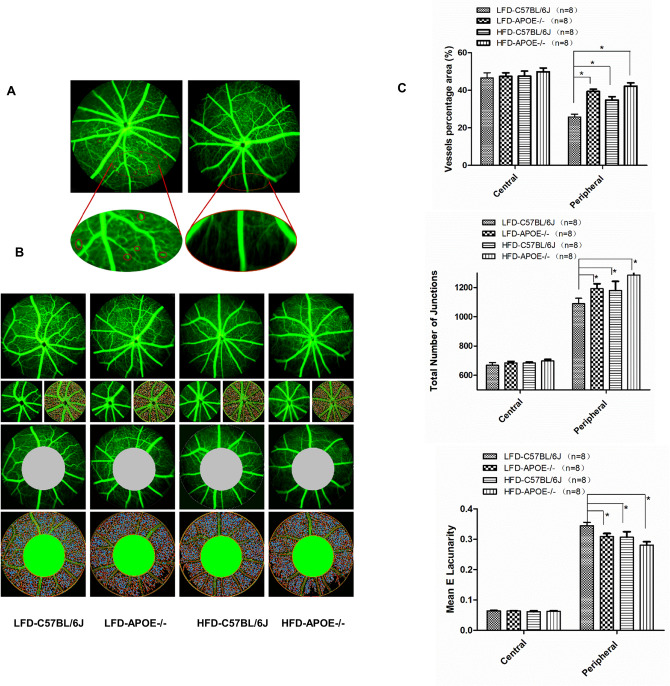
Figure 6.
Quantification of the capillary density. A HFD Apoe−/− mice exhibit microaneurysm and capillary non-perfusion zone in peripheral retina. B Microvessel density was assessed in the central and peripheral retina. C No statistically significant differences among four groups was observed comparing the central concerning vessels density, number of junctions and Mean E Lacunaroty. However, comparing the same values in the peripheral retina we observed significant differences among four groups. Data were shown as mean ± SEM. (n = 8). *P < 0.05.

