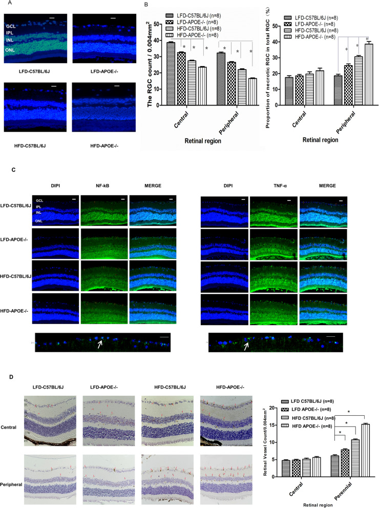
Figure 8.
HFD or apoE deficiency alone induced apoptosis of RGCs and vascular angiogenesis in retina. A HFD and apoE deletion decreased the density of Hoechst 33258-positive RGCs in the retina. Rats retinas from different groups were harvested at 32nd and subjected to whole mount immunostaining. Representative images from control, LFD-APOE−/−, HFD-C57BL/6J, HFD-APOE−/− group were stained by Hoechst 33258 (blue). Scale bar = 100 μm. B Densitometric analysis of the survival of the RGCs (Left) and proportion of necrotic RGCs (Right) in the GCL, which was assessed by counting the number of fluorescent Hoechst 33258 stained RGCs. Data were shown as mean ± SEM (n = 8, per group, *P < 0.01). C HFD and apoE deletion induced the activity of NF-κB and TNF-α in the peripheral areas in the retinal ganglion cells layer (GCL). Mice retinas from control, LFD-APOE−/−, HFD-C57BL/6J, HFD-APOE−/− group were stained by anti-NF-κB (green), anti-TNF (green) and DAPI (blue) Scale bar = 100 μm; Representative images from HFD-APOE−/− group (below), Scale bar = 50 μm. D HFD and apoE deletion increased the expression of VERGR2 in the peripheral areas in the retinal ganglion cells layer (GCL). The expression of VERGR2 was analyzed by H&E staining. Data of bar graph showed that compared with the VERGR2 expression in central area, a significant changes in the peripheral areas, especially in HFD-ApoE−/− group. The results were mean ± SEM. ***P < 0.001 (n = 8). Scale bar: 500 μm. GCL ganglion cell layer, IPL inner plexiform layer, INL inner nuclear layer, OPL outer plexiform layer, ONL outer nuclear layer, CHO choroid.

