
Fig. 3. EV assays with DMC and SEC samples.
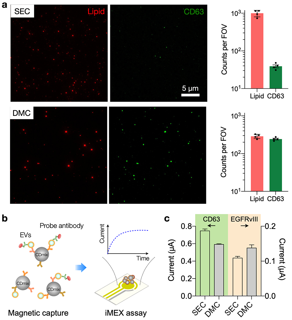
(a) Single particle imaging. SEC and DMC samples were labeled with the general lipid dye (red) and fluorescent CD63 antibodies (green). The SEC sample contained a large number of lipid particles per field-of-view (FOV, 77 × 65 µm2), but few of them were CD63-positive (top row). The DMC sample, in contrast, was enriched with CD63-positive lipid particles (bottom row). The graphs show particle counts (mean ± s.d.) from four FOVs. (b) Integrated magneto-electrochemical exosome (iMEX) EV protein assay. EVs are captured on magnetic beads based on EV-specific surface markers (CD63, CD81, CD9) and further labeled with probe antibodies to detect target protein markers. Probe antibodies, conjugated with oxidizing enzymes, generate electrical currents through electrochemical reaction. (c) Human plasma was spiked with EVs from Gli36 EFGRvIII mutation. Following SEC or DMC preparation, samples were assessed for CD63 and EGFRvIII expression via iMEX. The DMC sample showed a slightly lower CD63 signal than SEC, reflecting lower EV recovery. EGFRvIII signal, however, was higher in the DMC sample, presumably due to reduced interference from biological background. The data are from technical duplicates and displayed as mean ± s.d.