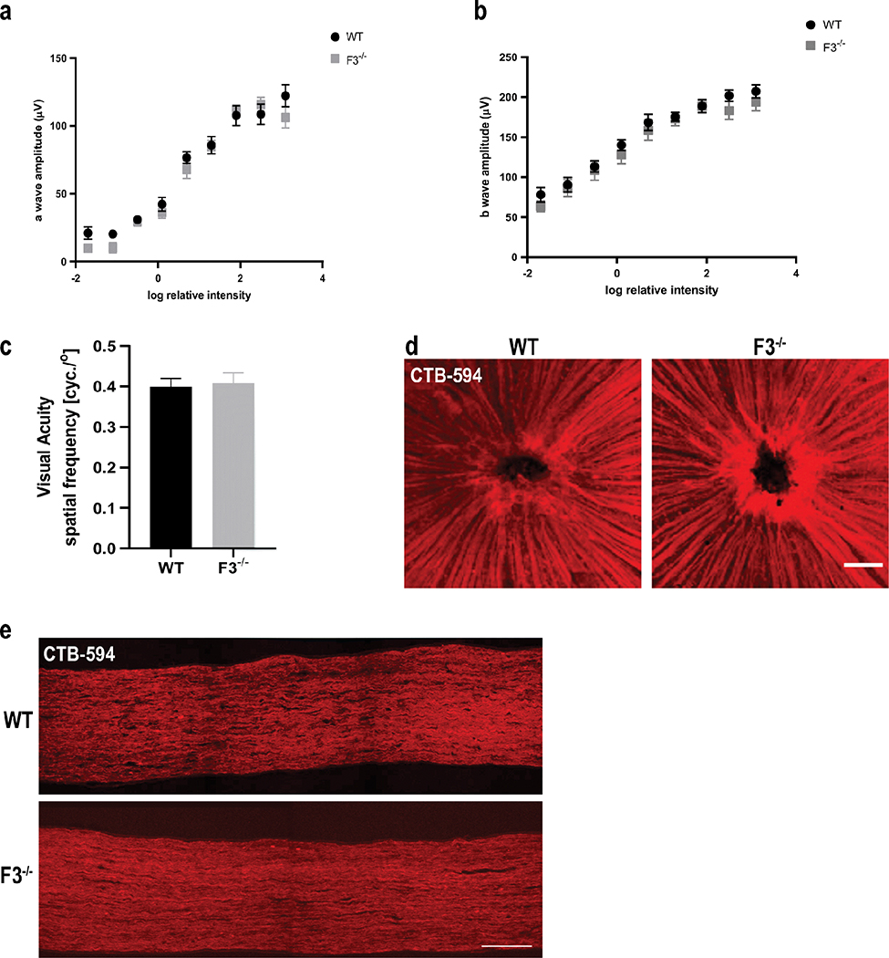
Fig. 6.
Normal retinal physiology and function are observed in the absence of F3. (a) An ERG exam revealed no significant differences in a-wave response (b) or b-wave response between WT and F3−/− animals at 6 months (n = 10–12 eyes). (c) Normal visual acuity was recorded for both WT and F3−/− animals by OKR at 2 months (n = 5). Analyses performed by 2-way ANOVA and represented as mean ± SD. (d) Representative images of CTB-594 tracer through nerve fiber layer (scale bar = 100 μm) and (e) optic nerve axons did not show any transport deficit in F3−/− mice compared with WT at 15 months (scale bar = 100 μm)

