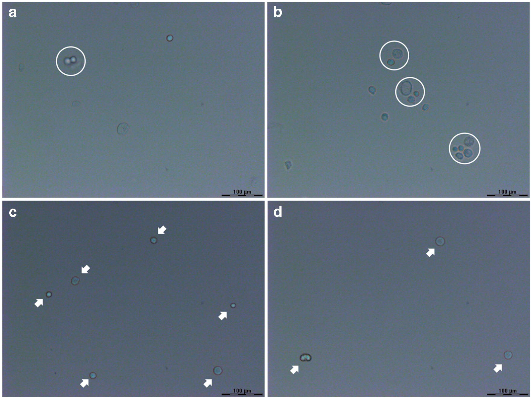
Figure 2.

Optical images of HeLa cells dispersed on the glass slides from (a, b) the original cell suspension and (c, d) the 10-fold dilution. The cell clusters in (a, b) are indicated by white circles, and the isolated single cells in (c, d) are pointed by white arrows. Scale bar = 100 μm.
