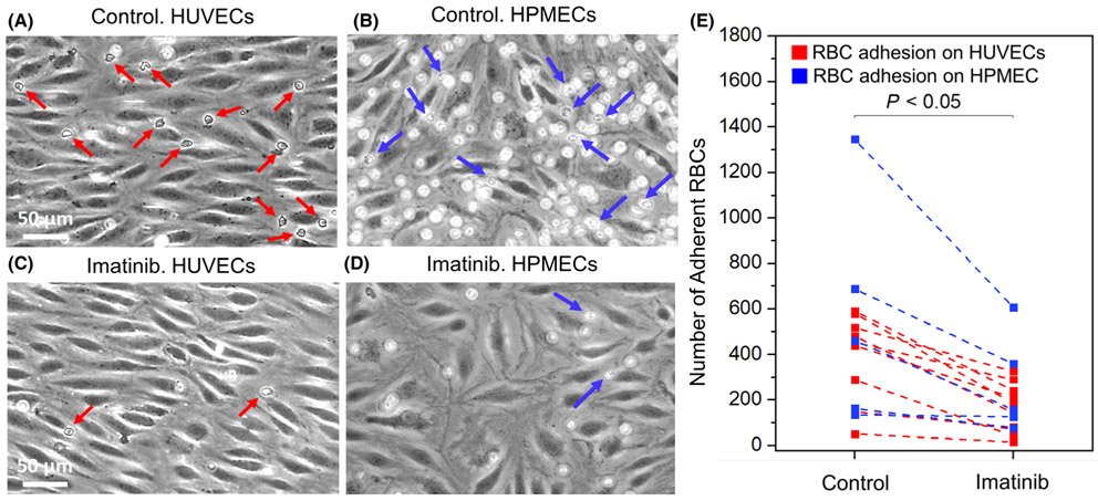
Fig 6.
Sickle RBC adhesion to heme-activated endothelial cells under physiologic hypoxia in microfluidic channels in vitro. Representative images of adherent RBCs to heme-activated endothelial cells are also shown in the control group (A, B on HUVECs and HPMECs) and in the imatinib-treated group (C, D on HUVECs and HPMECs). Arrows indicate RBCs adherent to endothelium. (E) Sickle RBC adhesion to heme-activated endothelial cells is significantly reduced by imatinib (5 μM) treatment, compared with control (vehicle, DMSO) treatment (N = 13 subjects, mean adhesion of untreated vs. imatinib-treated sickle cells ± SEM = 383 ± 57 vs. 171 ± 30, P < 0·001, paired t-test).). (RBC, red blood cell; HUVEC, human umbilical vein endothelial cells; HPMEC, human pulmonary microvascular endothelial cells).

