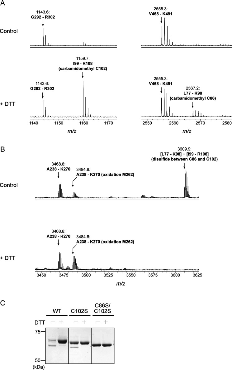
Figure 5.
Identification of redox-active Cys residues in Arabidopsis PGDH1. A and B, peptide mapping analysis based on MS. Before the Cys alkylation and following in-gel digestion with trypsin, the protein sample was incubated in the absence (control) or presence of 10 mm DTT. Overall mass spectra are shown in Fig. S4. C, redox shift assay of PGDH1 WT and Cys-substituted mutants (C102S and C86S/C102S) using the thiol-modifying reagent. Each PGDH1 was incubated with or without 10 mm DTT for 15 min. PGDH was then labeled with the methyl-PEG24-maleimide and loaded on nonreducing SDS-PAGE.

