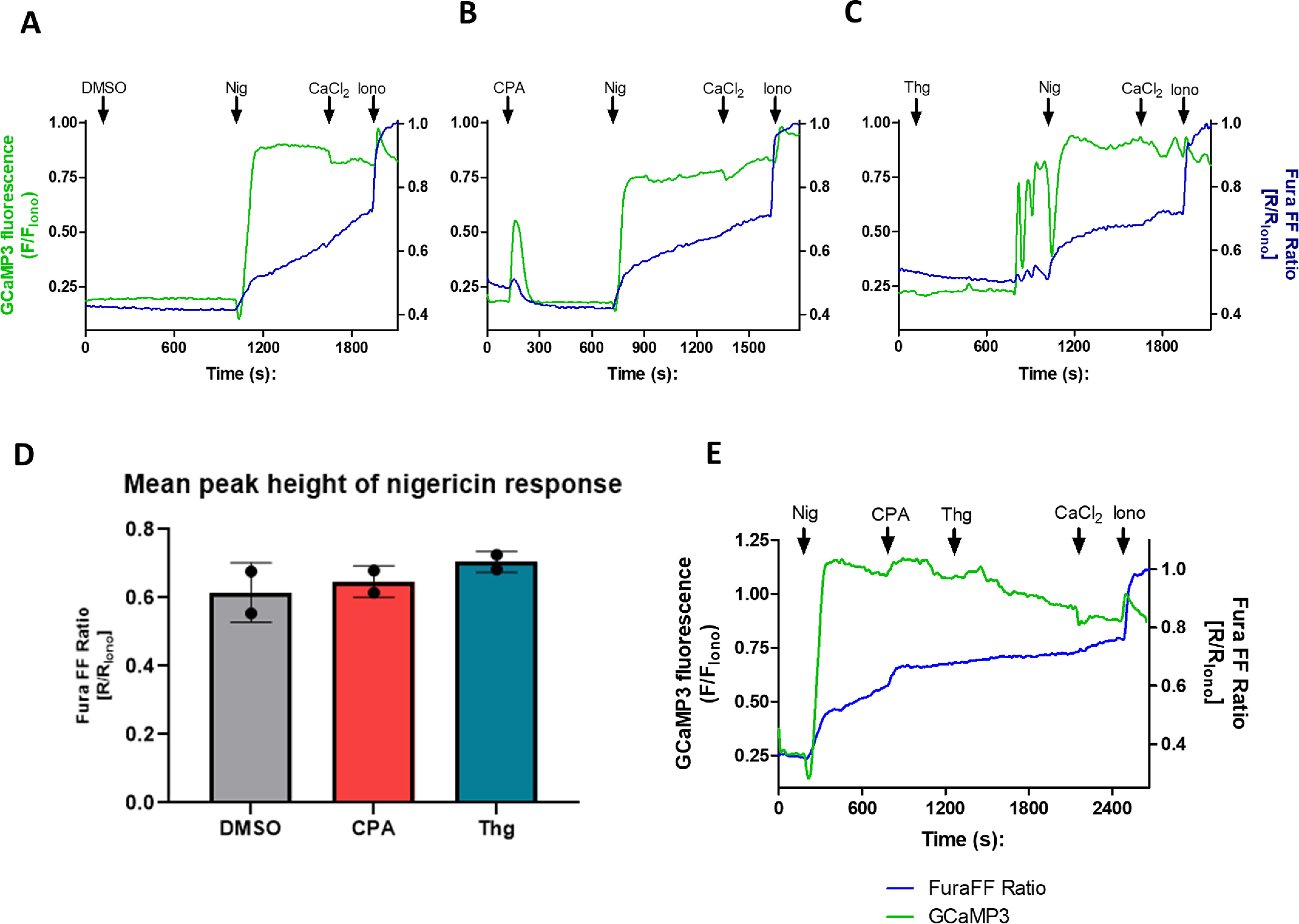
Figure 6.

The major acidic compartment is not affected by Thg or CPA. Isolated PfGCaMP3 parasites synchronized at the trophozoite stage were loaded with 5 μm Fura-FF/AM for 1 h while plating onto glass coverslips. The dual-loaded cells were transferred to the microscope imaging chamber, and immediately prior to recording, the buffer was switched to Ca2+-free MOPS buffer with 100 μm EGTA. The images were acquired at 1 Hz for both GCaMP3 (excitation, 488 nm; emission, 510 nm) and Fura-FF (excitation, 340 and 380 nm; emission, 510 nm). The cells were treated with either the DMSO vehicle control (A), 10 μm CPA (B), or 5 μm Thg (C) for 10–15 min, and then Nig (250 nm) was added for another 10 min prior to adding back 2 mm CaCl2 followed by 10 μm ionomycin (Iono). GCaMP3 fluorescence and the Fura-FF 340 nm/380 nm fluorescence ratio were normalized to the peak ionomycin (10 μm) response. The mean peak height of the Nig Ca2+ response was unaffected by CPA or Thg (D). Nigericin does not prevent a subsequent response to CPA and Thg (E). The data are the means ± S.D. of ≥60 cells from two independent experiments.
