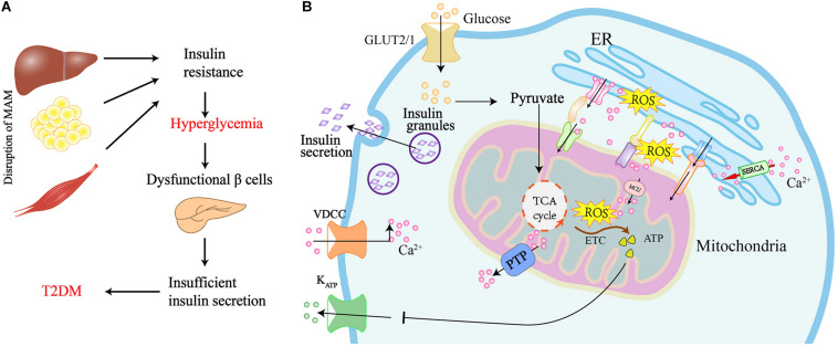
FIGURE 3.
Graphical summary of the MAM implicated in the insulin resistance of peripheral tissues and insulin secretion in pancreatic β cells. (A) Disruption of MAM leads to insulin resistance and the development of T2DM. (B) Scheme of MAM involved in the regulation of insulin secretion in pancreatic β cells. Glucose uptake via glucose transporter-2 (GLUT2) boosts mitochondrial ATP production and ROS generation by mitochondrial electron transport chain (ETC). Elevated ATP leads to the closure of ATP sensitive K+ channels (KATP) and the activation of voltage-dependent Ca2+ channels (VDCC), which ultimately results in the exocytosis of insulin-containing granules. Ca2+ flux into mitochondria via the MAM tethering complex increases ROS generation, which further promotes Ca2 + flux to the mitochondrial matrix by oxidizing the mitochondrial Ca2+ uniporter (MCU). The translocation of ROS from cristae to MAM results in the generation of redox nanodomains at the mitochondria-ER interface.

