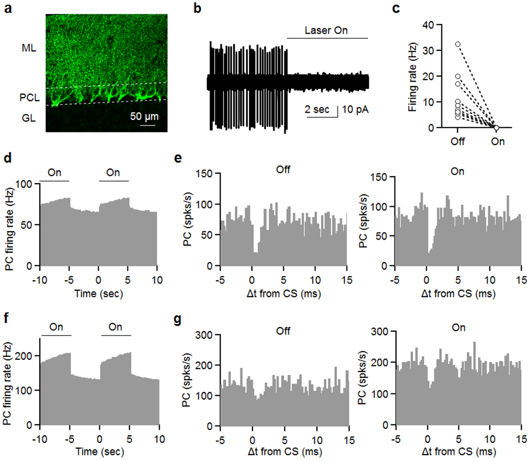
Extended Data Fig. 3. Optogenetic silencing of MLIs does not disrupt CS suppression of SSs in neighboring cells.

CKit cre mice53 were injected with 250 nl of AAV9-Ef1a-DIO eNpHR 3.0-EYFP nine sites in the cerebellum. Experiments were conducted 2-3 weeks later (see Methods). Slices were cut in order to evaluate expression and or ability to suppress MLI firing (a, b).
(a) Image of eNpHR 3.0-EYFP labelling showing an expression pattern that is characteristic of membrane labelling of MLIs, including the pinceaux associated with basket cells. 3 times reproduced.
(b, c) On cell recordings were used to assess the effect of light on MLI firing, and we found that firing was eliminated in all MLIs.
After waiting for 2-3 weeks, in vivo recordings proceeded similarly to experiments shown in Fig. 1. Once a pair was located, 5 s illumination was alternated with 5 s of no light, and this continued for at least 30 minutes in order to record sufficient complex spikes for each condition.
(d) Light increased PC firing of a pair of closely spaced (25 μm) cells.
(e) CS induced decreases in SS firing rate are shown for control condition (no light, left) and when MLI firing was suppressed with light (right).
(f) The effects of light on PC firing is shown for another pair of cells (50 μm).
(g) CS induced decreases in SS firing rate are shown for control condition (no light, left) and when MLI firing was suppressed with light (right).
