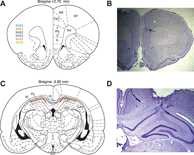
Figure 2 :
Electrode recording locations and histology. (A) Silicon probe recording locations in dmPFC for each rat. (B) Example photo of a cresyl violet stained coronal slice through PFC, showing the electrolytic lesion created to mark the recording location. (C) Tetrode recording region (highlighted in orange) was in the pyramidal layer of CA1 of dorsal HPC. (D) Example photo of a cresyl violet stained coronal slice through HPC, showing an electrolytic lesion and electrode track. Anatomy diagrams in (A) and (C) are from Paxinos and Watson (2006). IL, infralimbic cortex. PrL, prelimbic cortex. Cg1, cingulate cortex area 1. M2, secondary motor cortex. M1, primary motor cortex. fmi, forceps minor of the corpus callosum. hf, hippocampal fissure. Py, pyramidal layer. cc, corpus callosum. DG, dentate gyrus.

