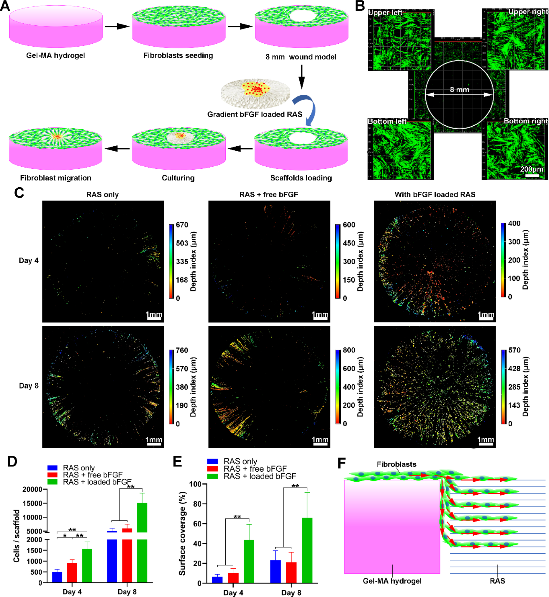
Figure 5. Applications of 3D radially aligned nanofiber scaffolds with a bFGF gradient along the radial direction for wound healing by recruiting cells towards the center.

(A) Schematic illustrating the 3D scaffold (8 mm) consisting of radially aligned nanofibers with gradations in the bFGF content that enhances wound healing by guiding and accelerating cell migration. The in vitro wound model (8 mm) is created by punching the Gel-MA hydrogel with GFP-labeled human dermal fibroblasts seeded on the surrounding area. (B) Fluorescent images show GFP-labeled human dermal fibroblasts around the 8-mm hole. (C) Fluorescent images show the migrated GFP-labeled human dermal fibroblasts into 3D scaffolds after 4 and 8 days of culture. (D, E) The quantification of cell counts and surface coverage (%) per scaffold after 4 and 8 days of culture. (F) Schematic illustrating the possible migration routes of GFP-labeled human dermal fibroblasts from the surface of the Gel-MA hydrogel to 3D scaffolds. *p<0.05, **p<0.01. RAS: radially aligned nanofiber scaffolds.
