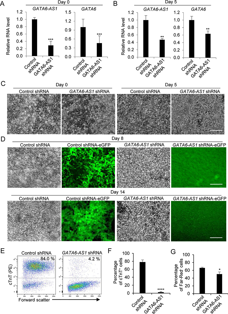
Figure 2. Knockdown of GATA6-AS1 inhibits the differentiation of cardiomyocytes induced by growth factors.
Control shRNA or GATA6-AS1 shRNA NKX2–5-eGFP hESCs were induced for cardiomyocyte differentiation using growth factors activin A and BMP4. (A, B) qRT-PCR analysis of GATA6-AS1 and GATA6 expression at day 0 and day 5. Data are normalized with amount of GAPDH mRNA relative to the corresponding value for cells transduced with the control shRNA. The mean fold difference and standard deviation in expression were calculated using three replicates. (C) Cell morphology of the control shRNA culture and the GATA6-AS1 shRNA culture during cardiomyocyte differentiation at day 0 and day 5. Scale bars = 200 μm. (D) Cell morphology and expression of NKX2–5-eGFP in the control shRNA culture and the GATA6-AS1 shRNA culture during cardiomyocyte differentiation at day 8 and day 14. Scale bars = 200 μm. (E) Flow cytometry analysis of differentiation cultures detected by α-actinin at day 14 in the control shRNA culture and the GATA6-AS1 shRNA culture. (F) Summary of the purity of cardiomyocytes from triplicate wells of the control shRNA culture and the GATA6-AS1 shRNA culture. (G) Summary of the viability of cardiomyocytes from triplicate wells of the control shRNA culture and the GATA6-AS1 shRNA culture. *, P<0.05; **, P<0.01; ***, P<0.001; ****, P<0.0001.

