Figure 2.
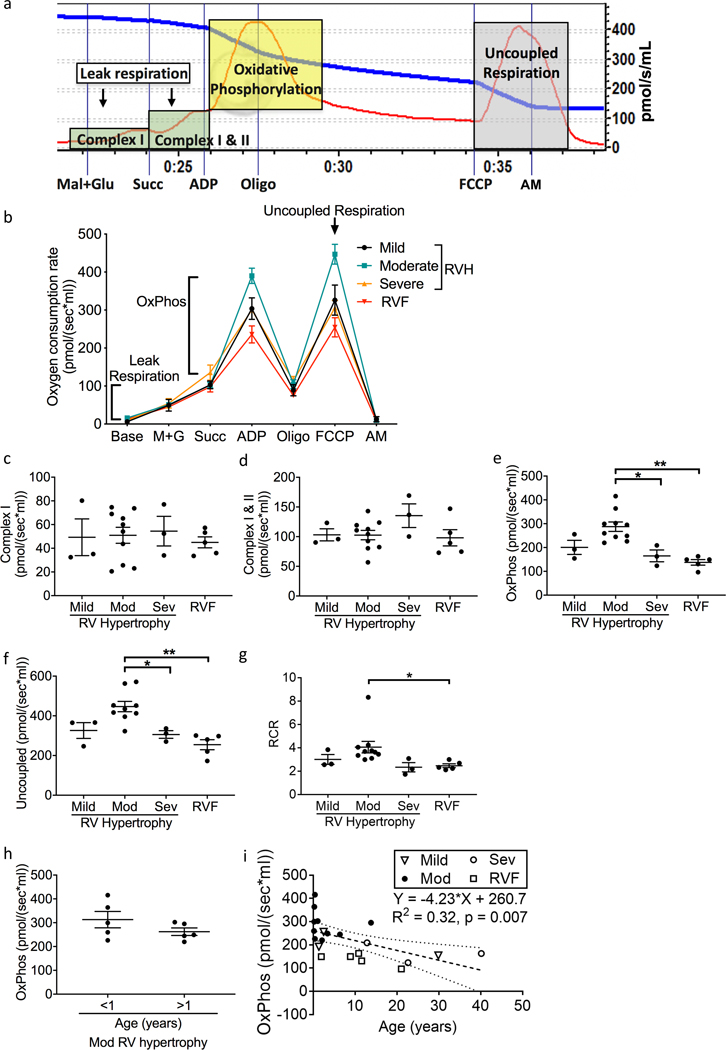
Patients with severe RV hypertrophy and RV failure demonstrate decreased oxidative phosphorylation. (a) A representative oxygen consumption tracing is shown for myocardial tissue (red curve) along with the available oxygen in the assay chamber (blue curve). We evaluated leak respiration (green shaded zones), oxidative phosphorylation (yellow shaded zone), and uncoupled respiration (gray shaded zone). (b) Summary of oxygen consumption in right ventricular myocardial tissue (N=3–10/group). (c, d) Complex I and Complex II mediated oxygen consumption was unchanged across all groups. (e, f) Oxidative phosphorylation and uncoupled respiration trended toward an increase from mild to moderate RV hypertrophy, decreased from moderate RV hypertrophy to severe hypertrophy, and decreased with RV failure. Oxidative phosphorylation remained significantly decreased after adjusting for age (p=0.0004) in RV failure. (g) Similarly, respiratory control ratio trended toward an increase in moderate hypertrophy and subsequently significantly decreased from moderate RV hypertrophy to RV failure. (h) Oxidative phosphorylation did not differ in patients with moderate RV hypertrophy between those <1 year of age versus >1 year of age (N=5/group). (i) Linear regression analysis demonstrates that age plays a role in the changes seen in oxidative phosphorylation but the actual change in oxidative phosphorylation per year (Slope) is small at −4.23pmol/sec*ml (N=21). RV – right ventricle; RVF – right ventricular failure; M+G – malate and glutamate; Succ – succinate; Oligo – oligomycin; FCCP - carbonyl cyanide-p-trifluoromethoxyphenylhydrazone; AM – antimycin A; Mod – moderate; Sev – severe; OxPhos - oxidative phosphorylation; RCR – respiratory control ratio. Data are presented as mean±SEM. *p<0.05, **p<0.01.

