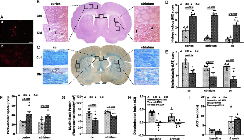
Fig. 1,
LD ME prompted histopathological damage in the brain of male diabetic animals. Fluorescence dye labeled ME were detected in petri dish (a in A) and in brain sections (b in A) at 3 days after injection (bar = 50 μm). The histopathological damage in the brain sections were analyzed with HE (B) and LFB (C) staining. Diabetic male animals with LD ME had more neuronal injury and vacuolation (arrow) in the cortex indicated by the higher HE score (D), lower LFB score (E), higher PVS index (F), and lower MBP intensity (G) in multiple brain areas indicating a greater degree of cerebral damage and demyelination compared to control animals. Diabetic male rats also had significantly lower discrimination index (d2) at baseline and 6 weeks after LD ME (H). However, the LD ME did not exacerbate cognitive deficits (H) and sensorimotor function in diabetic male rats (I). Ctrl: control, DM: diabetes, C-M: control male, D-M: diabetic male, cc: corpus callosum, ec: external capsule.

