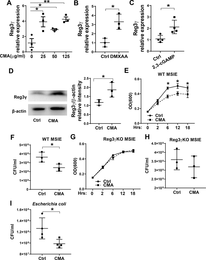
Figure 3. STING promotes IEC inhibition of bacterial growth through promoting REG3γ expression.
(A) MSIE cells (n = 3 /group) were treated with a series of doses of CMA for 24 hours. The expression of REG3γ was determined by qRT-PCR and normalized against GAPDH. (B) MSIE cells were treated with STING agonist DMXAA (1 μg/ml) for 24 hours, and the expression of REG3γ was determined by qRT-PCR and normalized against GAPDH. (C) MSIE cells were transfected with 2,3-cGAMP (1 μg/ml) for 48 hours, and the expression of REG3γ was determined by qRT-PCR and normalized against GAPDH. (D) Primary IECs of WT mice were treated with CMA (50 μg/ml) for 12 hours. The expression of REG3γ was determined by Western-blot with β-actin as the loading control. (E-H) WT MSIE cells and REG3γ KO MSIE cells were treated with or without CMA (50 μg/ml) for 24 hrs. The crude extracts were prepared and cultured with Citrobacter rodentium. The OD values (600 nm) were measured at different time points with the extracts of WT (E) and REG3γ KO (G) MISE cells. Bacterial culture suspensions were colonized to solid culture plates for colony counting after treated with the extracts of WT (F) and REG3γ KO (H) MISE cells. (H) *p < 0.05, **p < 0.01. (I) WT MSIE cells were treated with or without CMA (50 μg/ml) for 24 hrs. The crude extracts were prepared and cultured with Escherichia coli HS, and bacterial culture suspensions were colonized to solid culture plates for colony counting overnight. Data are represented as means ± standard deviation. One representative of 3–5 experiments with similar results was shown.

