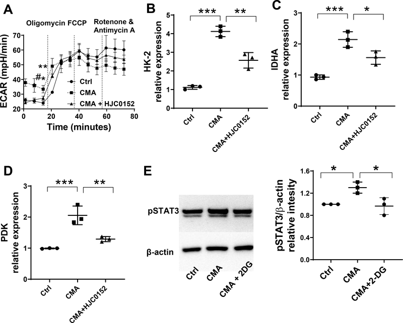
Figure 6. STAT3 and glycolysis interact each other in IECs upon STING stimulation.
(A) MSIE cells (n = 3/group) were treated with or without 50 μg/ml CMA in the presence or absence of STAT3 inhibitor HJC0152 for 24 hrs. ECAR was measured by Seahorse. (B-D) The expression of HK-2 (B), LDHA (C), and PDK (D) was determined by qRT-PCR and normalized against GAPDH. (E) MSIE cells (n = 3/group) were treated with or without 50 μg/ml CMA in the presence or absence of 2-DG (1 μM), and the protein levels of phosphorylated STAT3 were determined by Western blot. One representative of 2–3 experiments with similar results was shown.

