
FIGURE 6.
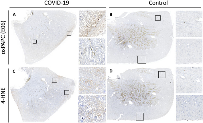
Immunohistochemical examination of renal tissue for potential ferroptosis markers. Renal tissue from the COVID‐19 patient with myocarditis and multiple organ dysfunction syndrome showed morphological signs of acute tubular necrosis, intratubular oxalate crystals, as well as E06 positivity in proximal tubuli (A). The latter also stained positively for the presence of 4‐HNE, one of the breakdown product of lipid peroxides (protocol adapted from Feng et al. 11 ) (C). By comparison, in the case of sudden death due to myocarditis of other aetiology, immunohistochemical staining with E06 (B) and anti‐4‐HNE antibody (D) in the renal tissue showed no presence of these ferroptosis markers (non‐specific staining in the corticomedullary junction is also present on control stains). Control stains using only secondary antibodies are enclosed in the Supporting Information.