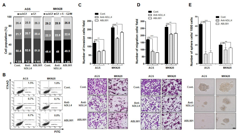
Fig. 2.
Cell cycle distribution, apoptosis, cell invasion, migration, and stem-like phenotype following treatment with anti-human DLL4 or ABL001 antibody in GC cells (A, B) AGS and MKN28 GC cells were treated with 50 nM anti-hDLL4 or ABL001 for 24 h. (A) Cell cycle distribution was detected by PI staining and FACS analysis. (B) Apoptosis induction was detected with Annexin V and 17-AAD staining and FACS analysis. (C, D) AGS and MKN28 GC cells were prepared and added to Transwell chambers. (C) The upper side of the membrane was coated with collagen type I for migration assay. (D) The lower side of the membrane was coated with Matrigel for invasion assay. After incubation with 50 nM anti-hDLL4 or ABL001 for 24 h, the migrated or invaded cells in the lower chamber were quantified following H&E staining by counting in five randomly selected areas in each well using wide-field microscopy. Data are expressed as mean ± standard error of mean (SEM) from three independent experiments. (E) AGS and MKN28 cells (1,000 cells each) were grown in ultra-low attachment plates containing mammary epithelium basal medium supplemented with or without anti-hDLL4 or ABL001. After culturing for 15 d, spheres with diameters of > 50 µm were counted. Cells were treated every 3 days with 50 nM concentration of the antibodies. *P < 0.05, **P < 0.01, and *** P < 0.001.

