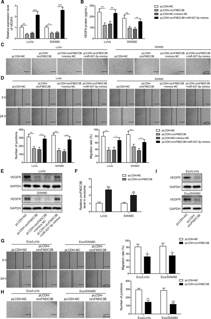
Fig. 4.

Overexpression of circFNDC3B suppressed CRC angiogenesis, which was reversed by miR‐937‐5p overexpression. (A, B) LoVo and SW480 cells were treated with pLCDH‐circFNDC3B alone or co‐transfected with miR‐937‐5p mimics, and the expression of VEGFA at mRNA and protein levels were determined by Q‐PCR (A) and ELISA (B), respectively. (C) HUVEC cells were treated with the CM collected from the cells above and the tube formation was observed under the microscope. Scale bar: 200 μm. (D) Changes of HUVEC migration abilities that were induced by the specific CM were assessed by wound‐healing experiment. Scale bar: 500 μm. (E) Western blot results showed that VEGFR expression in HUVEC was altered upon treatment of the indicated CM. (F) LoVo and SW480 cells were transfected with pLCDH‐NC or pLCDH‐circFNDC38, and the relative expression of circFNDC38 in exosomes was quantified by Q‐PCR. (G) Exosomes secreted by the cells in (F) were isolated and used to treat HUVEC. HUVEC migration was evaluated by a wound‐healing experiment upon treatment with the indicated exosomes. Scale bar: 500 μm. (H) Representative images of the tube formation in HUVEC upon treatment with the indicated exosomes are shown. Scale bar: 200 μm. (I) Western blot results showed that VEGFR expression in HUVEC was significantly decreased by treatment of the exosomes secreted by circFNDC3B overexpressing LoVo or SW480 cells. All the experiments were repeated three times. Statistical evaluation was performed using Student’s t test (two‐tailed) between two groups or one‐way ANOVA followed by Tukey’s post hoc test for multiple comparison. Data are expressed as mean ± SD. *P < 0.05; **P < 0.01; ***P < 0.001.
