
Fig. 3.
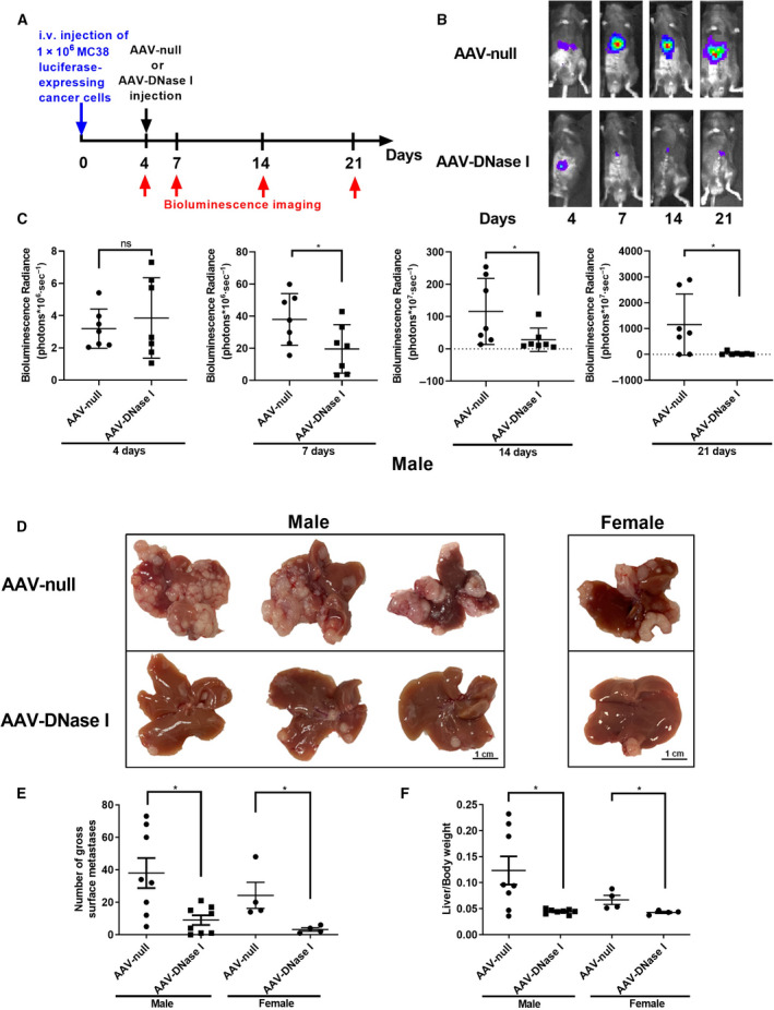
AAV‐mediated DNase I liver gene transfer reduces the growth of colorectal liver metastases. (A) Schematic showing experimental design in vivo. (B, C) The use of luciferase‐labeled MC38 cells allowed in vivo tracking of tumor growth with bioluminescence imaging. In the AAV‐DNase I‐treated group, we demonstrated that tumor growth is significantly inhibited at days 7, 14, and 21. n = 7 mice/group. Data were analyzed by Student's t‐test or Mann–Whitney U‐test. (D) Liver metastasis model using MC38 cells (1 × 10⁶) injected through the portal vein, showing smaller tumors harvested 3 weeks postinoculation in AAV‐DNase I‐treated mice compared with AAV‐null control. Scale bar = 1cm. (E, F) Graphs showing that AAV‐DNase I significantly decreased surface tumor nodules on liver and lower ratio of liver weight/body weight compared with AAV‐null group. n = 8 mice/group. Data were analyzed by Mann–Whitney U‐test. Data represent mean ± SD, *P < 0.05.