Fig. 4.

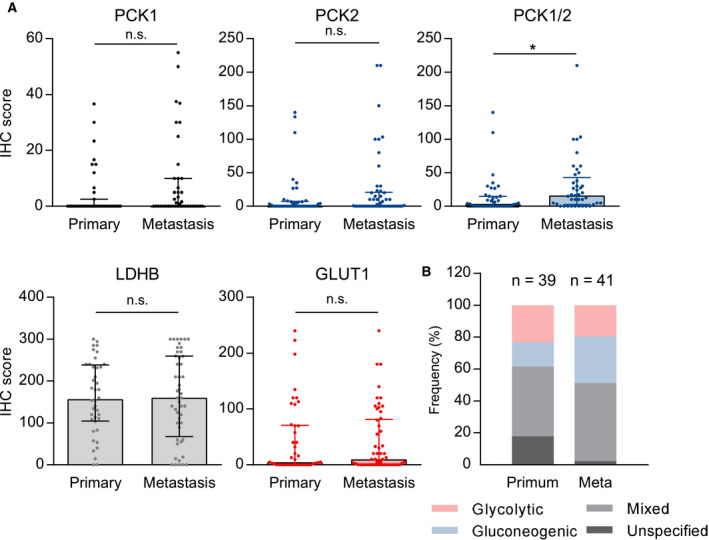
Gluconeogenesis and glycolysis enzyme expression in primary tumors and metastases. (A) A total number of 42 primary tumors from metastatic patients and 54 samples of NSCLC metastases were analyzed. No significant differences in IHC expression between primaries and metastases were observed in the separate analysis of PCK1, PCK2, LDHB, and GLUT1. The combined score of PCK1 and PCK2 (sum of the two scores) is significantly higher in the metastases. Group comparisons were performed with the Mann–Whitney U‐test in primary tumors and metastases with evaluable PCK1 (n = 39/41), PCK2 (n = 41/50), LDHB (n = 40/48), GLUT1 staining (n = 42/53), or PCK1/2 combined scores (n = 39/41). *P < 0.05. (B) distribution of glycolytic, gluconeogenic, and mixed phenotypes. Samples without immunopositivity for either PCK1, PCK2, or GLUT1 were defined as 'unspecified'.
