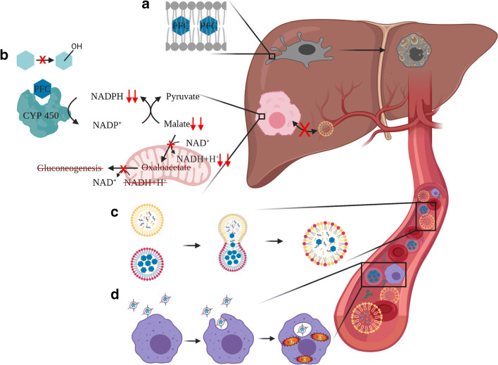
Fig. 2.
PFCs are inherently inert, but dependent on their lipo- and hydrophilicity and their vapour pressure they interact with the tissue and blood. (A) Some of the more lipophilic PFCs such as PFOB intercalate into the lipid bilayer of cell and organelle membranes [19]. The major part of PFCs are deposited inside the Kupffer cells which makes them appear foamy [84]. (B) PFCs can uncouple CYP450 monooxygenases, which reduces the detoxification capacity of the liver and causes differed glucose and lipid metabolism [21, 58, 71, 73, 85]. NAD/NADP: Nicotinamideadeninedinucleotide/-phosphate. (C) Proportional to the vapour pressure, phospholipid-wrapped PFCs (red) and natural lipid vesicles (yellow) inside the bloodstream may fuse and form hybrid vesicles [53, 99]. (D) PFC droplets are opsonized by complement factors or antibodies to be recognized and phagocytized by macrophages [52]. Figure created with Biorender.com

