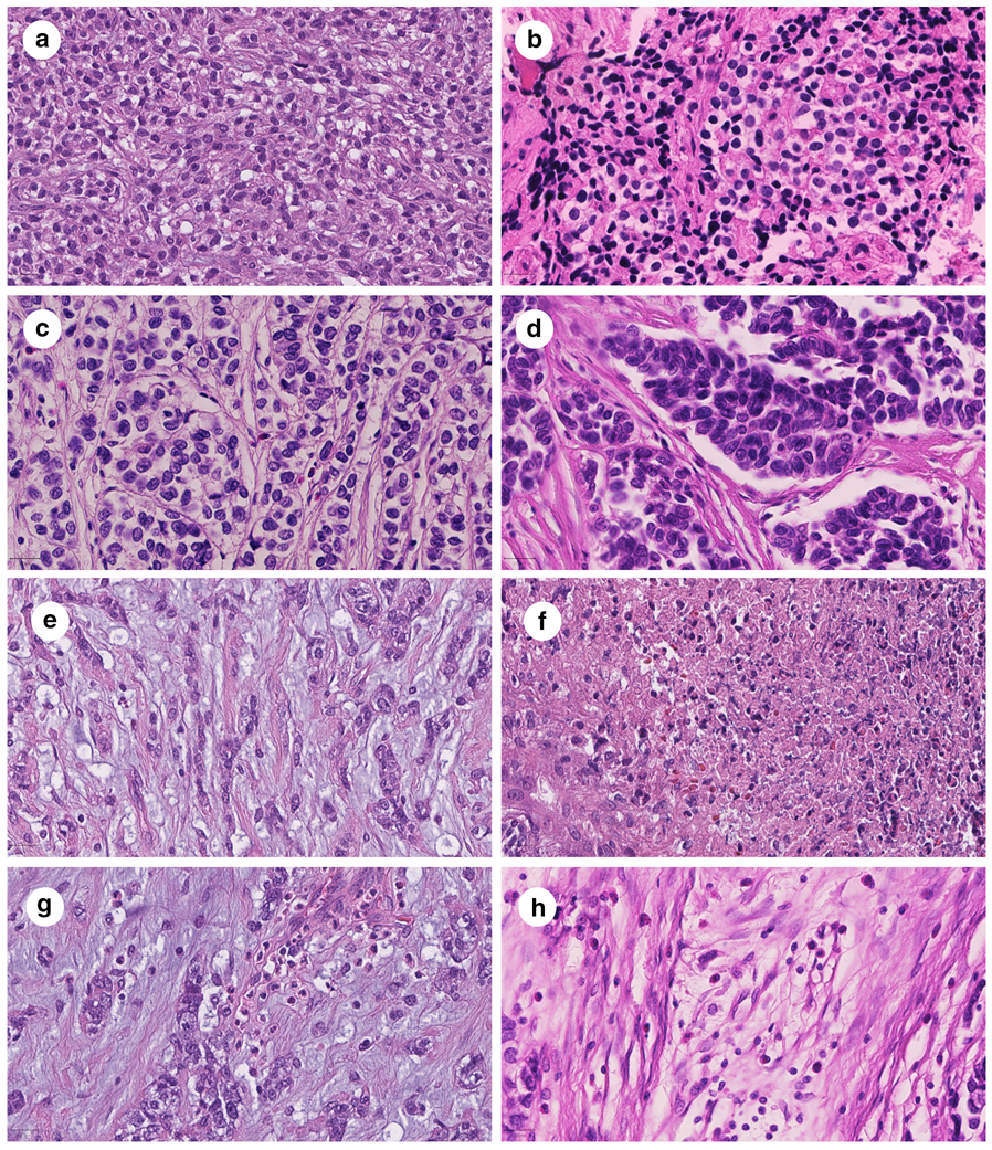
Figure 1: Histologic features of undifferentiated round cell sarcomas with EWSR1-NFATc2 fusion.

Undifferentiated round cell sarcomas with EWSR1-NFATc2 fusion were composed of relatively monotonous cells with a faint eosinophilic to clear cytoplasm and large, chromatin dense nuclei (a, b). The tumor matrix variably contained thin to coarse collagen bundles, which separated tumor cells and appeared as a nested growth pattern (c, d). One case presented with a myxoid tumor matrix (e). Focal necrosis was observed in two cases (f). Inflammatory cells were variably present and were composed of eosinophilic leukocytes (g, h). Magnification: 400-fold. Scale bars: 20 μm.
