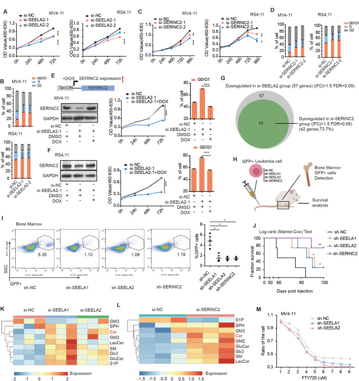
Fig. 4.
SEELA-SERINC2 axis promotes leukemia progression in vitro and in vivo through sphingolipid metabolism. a-d Inhibition of SEELA2 (a, b) and SERINC2 (c, d) disrupted the cell proliferation and induced the accumulation of G0/G1 phase cells via CCK-8 assay and cell cycle assay by flow cytometry in MV4–11 and RS4;11 cells. Error bars reflect ± SEM (*P < 0.05; **P < 0.01; ***P < 0.001) in three independent experiments. e, f Schematic outline of SERINC2 rescue strategy (e left upper) in MV4-11 (e) and RS4;11 (f) cells. Western blot showed the protein level of SERINC2 in indicated cells. CCK8 and cell cycle assay showed the cell proliferation and cell cycle arrest of indicated treatment. The error bars indicate the ± SEM values (***P < 0.001) in three independent experiments. g Total 57 genes were dysregulated in the si-SEELA group (|FC| > 1.5, FDR < 0.05), and 42 of 57 genes (73.7%) were dysregulated si-SERINC2 group (|FC| > 1.5, FDR < 0.05). h A schematic diagram shows the xenotransplantation model. i Representative flow cytometry graphs show the decreased percentages of human leukemic GFP+ cells in the bone marrow from mice treated with sh-SEELA, and sh-SERINC2 treated MV4-11 cells relative to the levels observed in control mice (left). A scatter plot shows the statistical values (right). Error bars reflect ± SEM (*P < 0.05). j Kaplan-Meier survival curves show that the mice injected with sh-SEELA and sh-SERINC2 survived longer than those of the control sh-NC group (n = 4). The P values were calculated using a log-rank (Mantel-Cox) test. (*P < 0.05; **P < 0.01). k, l Heatmaps show the expression of sphingolipids from the mass spectrometry (MS) assay. Most of the sphingolipids were upregulated after knocking down SEELA (si-SEELA1-1 or si-SEELA2-1) (k) or SERINC2 (si-SERINC2-1) (l) in MV4-11 cells. Ceramides, Cer; sphingomyelins, SM; glucosylceramides, GluCer; lactosylceramides, LacCer; ganglioside, GM2; monosialo-dihexosyl gangliosides, GM3; trihexosylceramide, Gb3; sphingosine, SPH; and sphingosine-1-phosphate, S1P. m The survival rate of MV4-11 cells was measured using a CCK-8 kit at 24 h. The sh-NC, sh-SEELA established cells were treated with FTY720 at concentrations from 1 to 9 μM

