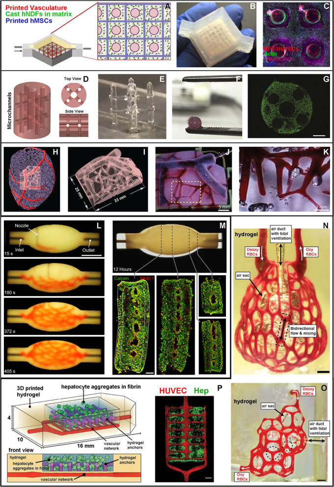
Fig. 1.
a-c “Thick perfusable construct” (adapted from Kolesky et al., 2016). a Schematic representation of the printed tissue composed of: i. sacrificial ink to fabricate the vasculature network (red circles), ii. human mesenchymal stem cell (hMSC) laden hydrogel (blue squares), and iii. The surrounding hydrogel containing human neonatal dermal fibroblasts (hNDF, green dots); b picture of the final bioreactor containing the tissue construct; c confocal image of a cross-section after 30 days of perfusion; the construct is densely populated by viable cells (hMSCs, DAPI and actin marking nuclei and cytoskeleton) with a higher osteocalcin expression the closer they are to the channels; HUVECs surrounding the internal cavity of the channels are also visible. d-g “Implantable vascularized construct for bone repair” (adapted from Daly et al., 2018). d Schematic representation of the vascularized construct; e freestanding filament network printed with PLU; f final GelMA construct after PLU wash out; g fluorescence image showing live/dead (green/red) MSCs 24 h after fabrication, scale bar 500 μm. H-K. “Multi-scale MRI-derived vascular network” (adapted from Lee et al., 2019). h Computational representation of left ventricle vasculature; i subregion chosen for 3D bioprinting; j perfusion of the final structure with magnified detail in k. l-m “Branched vascular network printed in a spheroid support bath” (adapted from Skylar-Scott et al., 2019) l images at different timepoints during printing of the branching vascular network in a spheroid-based matrix, scale bar 10 mm; m fluorescence images at different sections (dashed lines) after 12 h of perfusion, showing live/dead (green/red) iPSCs forming the spheroids. n-p “Vascularized alveolar and hepatic tissue models” (adapted from Grigoryan et al., 2019). n, o Printed alveolar models, characterized by a central air sac surrounded by a blood perfused vascular network, scale bars 1 mm; p schematic representation and fluorescence imaging of a hydrogel loaded with hepatic cells (green) and supported by a vascular network (red)

