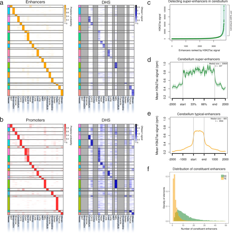
Fig. 1.
Overview of TSREs identified in 22 mouse tissues. a Strong enhancers, b Active promoters: Heatmaps showing chromatin state posterior probability of tissue-specific regulatory elements (Taureg ≥ 0.85) (left) and their corresponding DNAse1 signal (right) in every tissue. Each row is a genomic location and columns represent different mouse tissues and cell lines. Grey columns show tissues for which data was not available. The heatmaps have been sorted by the order of the tissues across the columns. (BAT: Brown Adipose Tissue; Bmarrrow: Bone Marrow; BmarrowDm: Bone Marrow derived macrophage; CH12: B-cell lymphoma; Esb4: mouse embryonic stem cells; Es-E14: mouse embryonic stem cell line embryonic day 14.5; MEF: Mouse Embryonic Fibroblast; MEL: Leukaemia; Wbrain: Whole Brain). c Distribution of H3K27ac ChIP-seq signal over cerebellum-specific enhancers stitched together within 12.5 kb (n = 3741). Stitched cohesive units (x-axis) are ranked in an increasing order of their input-normalised H3K27ac signal (reads per million, y-axis). This approach identified 237 SEs (highlighted in blue) and 3504 TEs in cerebellum. d-e Metagene profile of mean H3k27ac ChIP-seq signal across all the SEs and TEs in cerebellum. The profiles are centred on the enhancer regions and the surrounding 2 kb regions around each enhancer is shown. The length of the enhancer region is scaled to represent the median size of SEs (22,600 bp) and TEs (600 bp) in cerebellum. The shaded area shows the standard error (SEM). f Distribution of constituent enhancers within SEs and TEs across all 22 tissues. See also Additional file 1: Figure S2-S5

