Fig. 2.
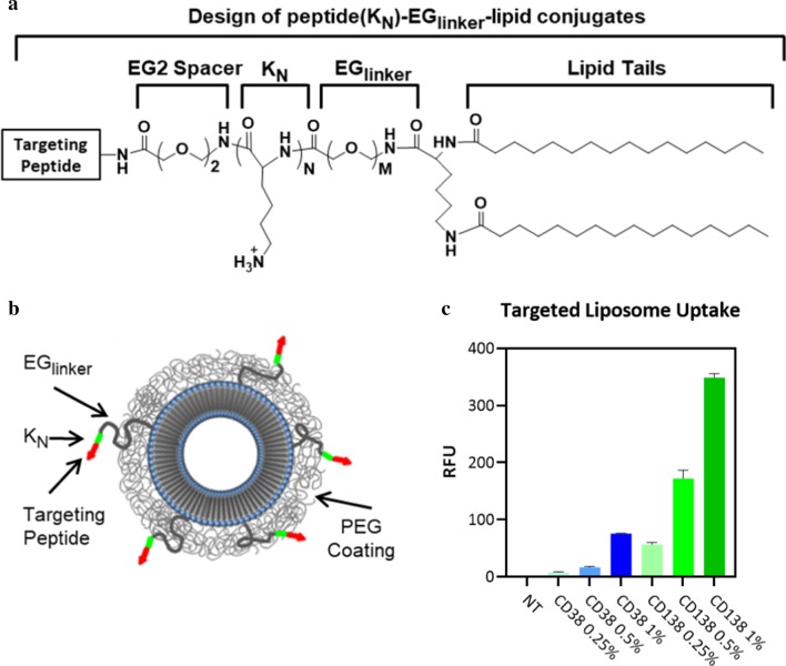
Design of peptide conjugated liposomal nanoparticles and uptake of targeted nanoparticles into H929 myeloma cells. a Design of peptide(KN)–EGlinker–lipid conjugates with variable oligolysine (KN) content and EG peptide–linker lengths. b Schematic of the peptide-targeted nanoparticles. c Uptake of nanoparticles targeted with varying densities of CD38pep or CD138pep. Nanoparticles were incubated with H929 cells in media for 3 h, trypsinized to remove nanoparticles bound to the surface but which had not yet undergone cellular uptake, and then fluorescence was measured by flow cytometry. All experiments were done in triplicate. Data represent means (± s.d.)

