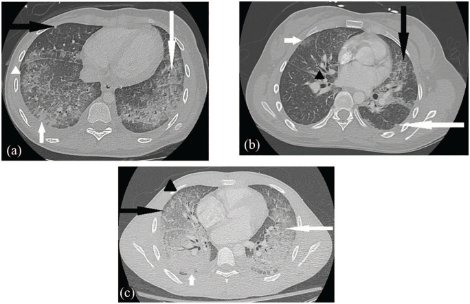
Figure 2.
Chest CT for case 1 (image a) demonstrates ground glass opacities (long white arrow), patchy consolidation (short white arrow), crazy paving appearance (white arrowhead) which describes ground glass opacities with septal thickening, nodular foci (black arrow), and subpleural sparing (also white arrowhead). Chest CT for case 2 (image b) displays scattered mostly peripheral ground glass lung opacities (short white arrow) with nodules (long white arrow), septal thickening (black arrow), and peribronchial wall thickening (black arrowhead). Chest CT for case 3 (image c) shows interlobular septal thickening (black arrowhead) and alveolar ground glass opacities (white arrow) throughout much of the lungs with notable subpleural sparing (black arrow), more consolidative opacities in dependent lung bases (small white arrow), and small bilateral pleural effusions.

