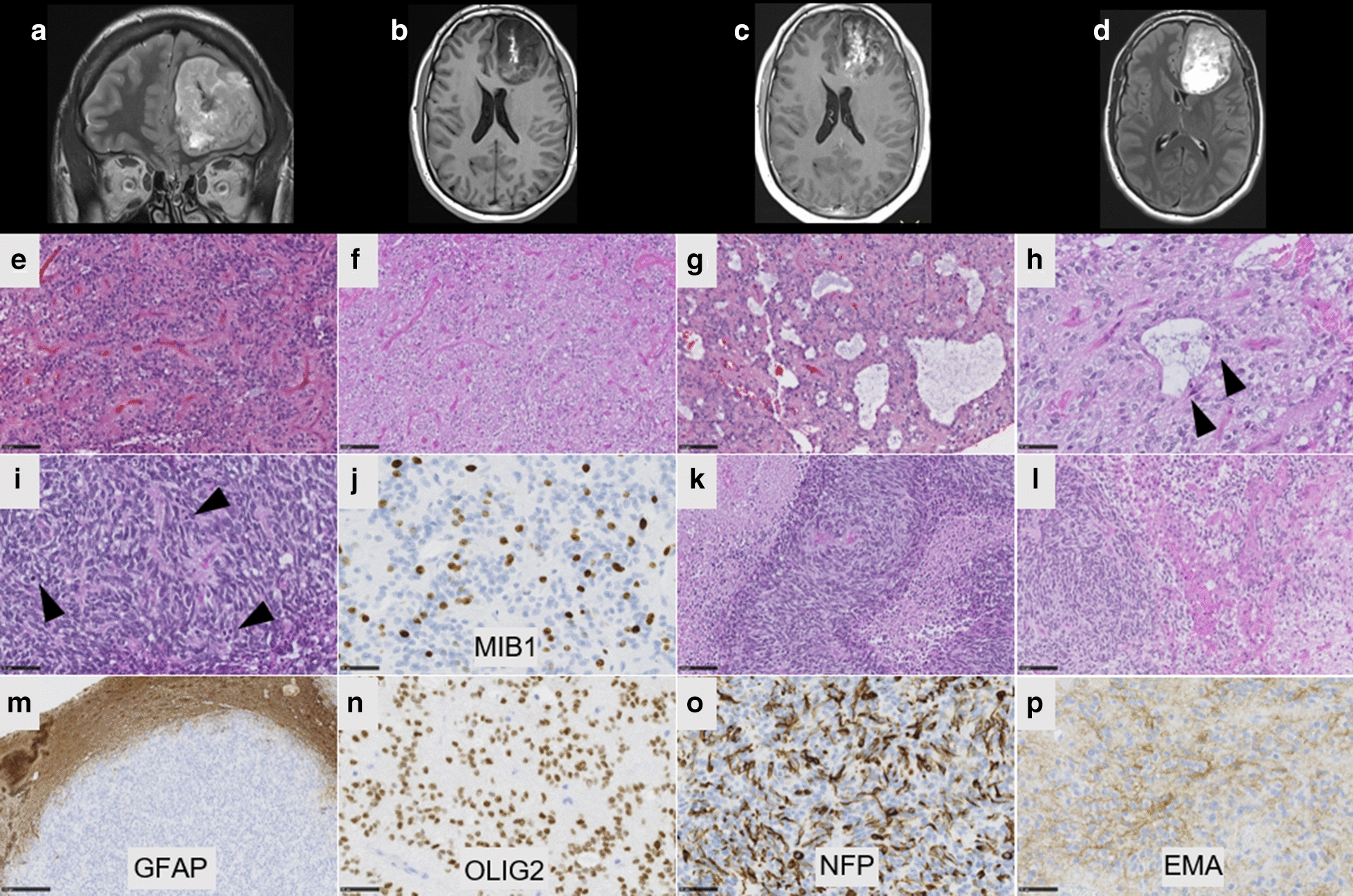
Fig. 2.

Radiological and histopathological features of #case 2. a Coronal T2-weighted sequence showing a large tumor without peri-lesional edema in the left frontal lobe. b Axial T1-weighted image showing a left frontal mass with leptomeningeal attachment and heterogeneous enhancement after gadolinium injection. c T1-weighted image after injection of gadolinium showing a heterogeneous enhancement. d Flair sequence showing hyperintensity. e Compact tumor with delicate branching vessels exhibiting a chicken-wire pattern (HPS, magnification ×200) with oligo-like features (f, HPS, magnification ×200). g Microcyst with a sometimes myxoid background (HPS, magnification ×200) and h containing some neuronal cells (arrowheads, HPS, magnification ×400). i Area with dense cellularity and high mitotic index (arrowheads, HPS, magnification ×400) and j elevated MIB1 labeling index (magnification ×400). k Palisading necrosis (HPS, magnification ×400) and microvascular proliferation (l, HPS, magnification ×400). m The tumor is well-circumscribed from brain parenchyma, as seen on GFAP staining, without expression in the tumor (magnification ×100). (n) Diffuse expression of Olig2 (magnification ×400). o Neurofilament expression by tumor cells (magnification ×400) and p cytoplasmic expression of EMA (magnification ×400). Black scale bars represent 100 µm (e–g, k,l), and 50 μm (h,i, n–p) and 250 µm (m)
