Fig. 3.
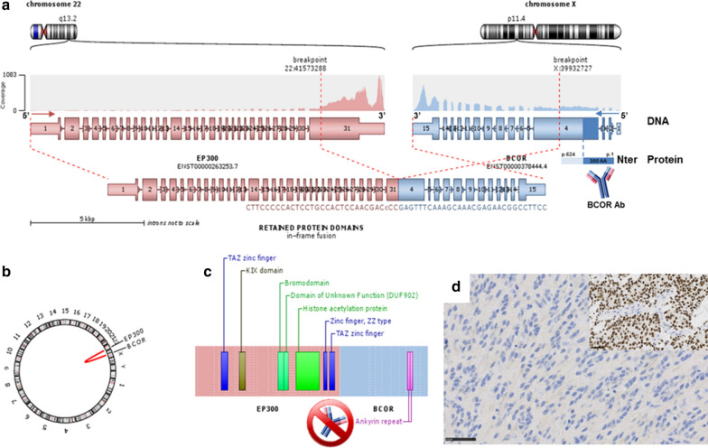
Fusion EP300:BCOR and correlation with immunohistochemistry. a RNAseq analysis highlights a fusion between EP300 (pink) and BCOR (blue) genes, respectively located on chr22q13.2 and chrXp11.4. As the breakpoints are intra exonic (in exon 31 for EP300, and exon 4 for BCOR), the fusion point can easily been detected by split and span reads encompassing the rearrangement with a good coverage. Localized on minus strand (inverse orientation), the DNA sequence of BCOR is switched in frame with EP300 (b Circos plot). This aberration causes the loss of the first 3 exons of BCOR and a part of the exon 4 encoding the Nter domain of the protein (dark blue). As the BCOR antibody is designed against the 300 first residues of the native protein and since this epitope is missing in the resulting chimeric fusion protein, it cannot be used for EP300-BCOR detection by IHC. c Conserved domains in the fusion protein. d Absence of expression of BCOR by immunohistochemistry with positive internal control (tumor of methylation class HGNET-BCOR with BCOR internal tandem duplication, insert) (magnification ×400). Black scale bars 50 μm (D)

