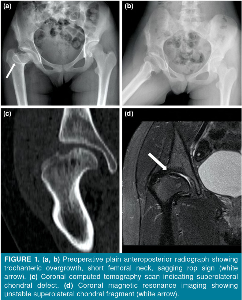
Figure 1. (a, b) Preoperative plain anteroposterior radiograph showing trochanteric overgrowth, short femoral neck, sagging rop sign (white arrow). (c) Coronal computed tomography scan indicating superolateral chondral defect. (d) Coronal magnetic resonance imaging showing unstable superolateral chondral fragment (white arrow).