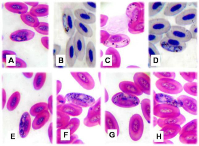
Fig. 2.
Microphotographs of Haemoproteus columbae (×1000). (A) Immature macrogametocyte, (B) Immature micro-gametocyte, (C) Mature microgametocyte broad at one end and narrow at the other, (D) Mature microgametocyte rounded at both poles, (E) Mature macrogametocyte encircled host cell nucleus, (F&G) Mature macrogametocyte displaced host cell nucleus to periphery, and (H) Extra-corpuscular form

