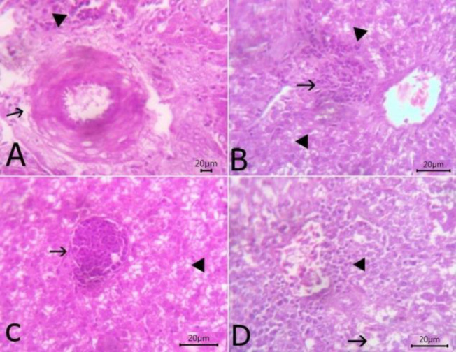
Fig. 3.
The histopathological lesions in liver sections stained with H&E. (A) The liver showing slight congestion of portal vein with thickening of the wall and perivascular edema (arrow) beside few mononuclear cells infiltration (arrowhead) (bare=20), (B) Liver showing vacuolar degeneration of hepatocytes (arrowhead) with interstitial aggregation of round cells and fibroblasts (arrow) (bare=20), (C) Liver showing round-shaped thin-walled megaloschizont of Haemoproteus columbae (arrow) and vacuolation of hepatocytes (arrowhead) (bare=20), and (D) Liver showing hydropic degeneration of hepatic cells (arrow) and congestion of central vein with a perivascular aggregation of mononuclear inflammatory cells (arrowhead) (bare=20)

