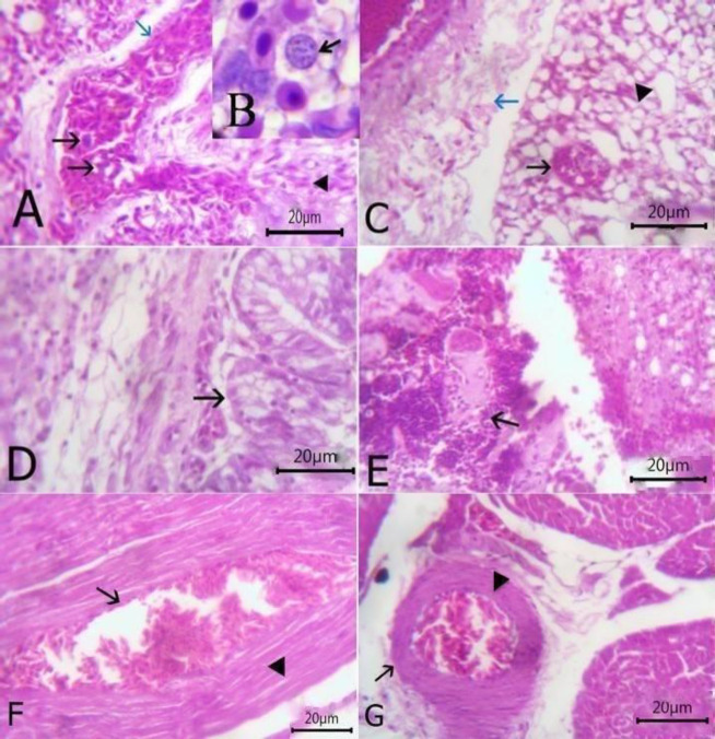
Fig. 4.
(A) Lung section stained with H&E showing the presence of Haemoproteus columbae schizont within pulmonary blood vessel (arrow), dilated and congested (blue arrow) with presence extravasated erythrocytes (arrowhead) (bare=20), (B) Higher magnification of schizont (arrow), (C) Lung showing schizont inside the alveoli (arrow) with some emphysematous alveoli (arrowhead) and perivascular edema around congested blood vessels (blue arrow) (bare=20), (D) Lung showing catarrhal bronchitis (arrow) (bare=20), (E) Lung showing granuloma which consisting of the central area of caseous necrosis surrounded by mononuclear inflammatory cells (arrow) (bare=20), (F) Heart showing extravasated erythrocytes (arrow) among degenerated cardiac muscles (arrowhead) (bare=20), and (G) Heart showing congestion of blood vessels with hyaline thickening of its wall (arrow) and perivascular edema (arrowhead) (bare=20)

