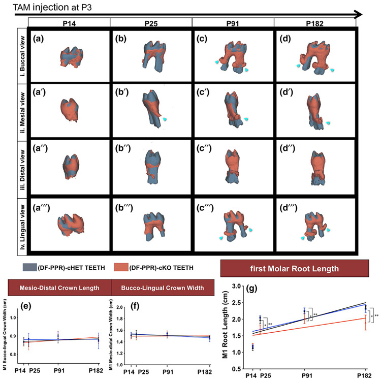
FIGURE 3.
Mouse primary failure of eruption (PFE) molars show shorter and dilacerated roots. (a-d") Composite 3D surface model overlay of the mandibular first molars (M1), views from the buccal (a-d), mesial (a'-d'), distal (a"-d") and lingual (a"'-d"') side (DF-PPR) cHet and (DF-PPR) cKO M1 were superimposed on the crown. Dark gray: (DF-PPR) cHet, red: (DF-PPR) cKO. Blue arrowheads: truncation (short roots) associated with dilacerations (curved roots). (e-g) Quantitative 3D-μCT analysis. M1 mesiodistal crown length (e), buccolingual crown width (f) and root length (g) measured on 3D-μCT images. Colored lines: regression line, black: Control, blue: (DF-PPR) cHet, red: (DF-PPR) cKO. At P14 n = 5, P25 n = 6, P91 and P182 n = 4 **p < .01, *p < .05, One-way ANOVA followed by Mann-Whitney's U test. All data are represented as mean ± SD

