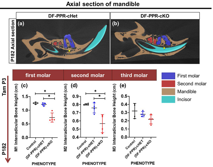
FIGURE 4.
Mouse primary failure of eruption (PFE) molars defective cryptal bone formation. (a,b) Axial sections of 3D-μCT images, (DF-PPR) cHet (a) and (DF-PPR) cKO (b) at P182. Blue: M1, red: M2, beige: mandible, blue: incisor. Arrowheads: interradicular cryptal bone. (c-e) Quantitative 3D-μCT analysis. Interradicular bone height of M1 (c), M2 (d) and M3 (e) measured on 3D-μCT images. *p < .05, One-way ANOVA followed by Mann-Whitney's U test. All data are represented as mean ± SD

