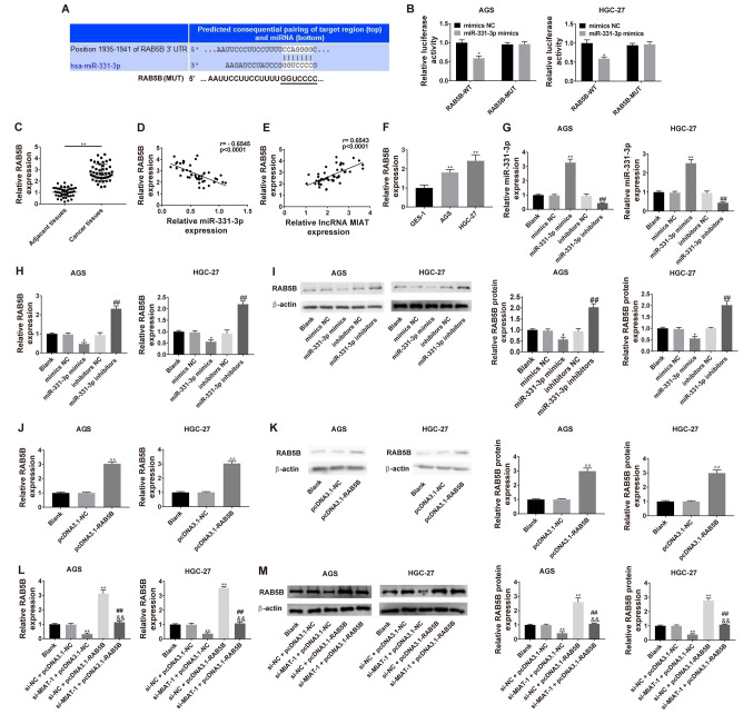
Figure 5.
Dual-luciferase reporter system detection of the effect of miR-331-3p on RAB5B in HGC-27 and AGS cells. (A) Predicted binding between miR-331-3p and RAB5B. (B) Luciferase activities of RAB5B-WT or RAB5B-MUT reporter were detected via dual-luciferase reporter assay. *P<0.05 vs. mimics NC/RAB5B-WT. (C) RAB5B levels were measured via RT-qPCR in 47 pairs of GC tissues and adjacent tissues. (D) Correlation analysis between miR-331-3p and RAB5B in 47 GC tissue samples. n=3. **P<0.01 vs. the adjacent tissues group. (E) Correlation analysis between MIAT and RAB5B in 47 GC tissue samples. (F) RAB5B levels were detected via RT-qPCR in different cell lines (GES-1, HGC-27 and AGS). n=3. **P<0.01 vs. the GES-1 group. (G) HGC-27 and AGS cells were transfected with mimics NC, miR-331-3p mimics, inhibitors NC or miR-331-3p inhibitors, followed by the measurement of miR-331-3p level. HGC-27 and AGS cells were transfected with mimics NC, miR-331-3p mimics, inhibitors NC or miR-331-3p inhibitors, followed by the measurement of RAB5B level at the (H) mRNA and (I) protein levels. n=3. *P<0.05 vs. the blank or mimics NC group; ##P<0.01 vs. the blank or inhibitors NC group. (J) HGC-27 and AGS cells were transfected with pcDNA3.1-NC or pcDNA3.1-NC, followed by (K) the measurement of RAB5B level. n=3. ^^P<0.01, compared with the blank or pcDNA3.1-NC group. (L) Transfection efficiency in co-transfected AGS and HGC-27 cells. (M) Measurement of RAB5B level at 48 h after co-transfection. n=3. *P<0.05, **P<0.01 vs. the blank or si-NC + pcDNA3.1-NC group; ##P<0.01 vs. the si-MIAT-1 + pcDNA3.1-NC group; &&P<0.01 vs. the si-NC + pcDNA3.1-RAB5B group. GC, gastric cancer; MIAT, myocardial infarction associated transcript; si, small interfering; NC, negative control; miR, micro RNA; WT, wild-type; MUT, mutant; RT-qPCR, reverse transcription-quantitative PCR.

