Fig. 5.
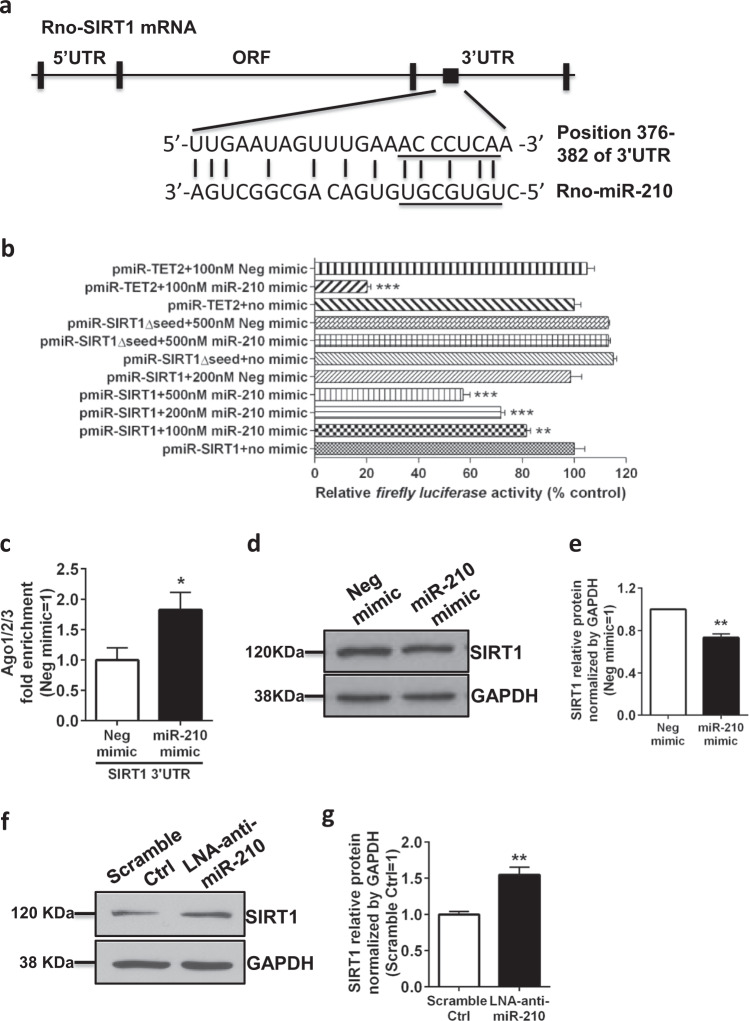
SIRT1 is a bona fide target of miR-210 in neonatal rat microglia. a. Schematic representation of alignment of the predicted miR-210 binding site to the SIRT1 3′UTR is shown for Rattus norvegicus. b The rat PC12 cell line was cotransfected with the indicated constructs (500 ng/well) containing the wild-type or mutant 3′UTR of SIRT1 or the wild-type 3′UTR of TET2 (positive control) in the presence of miR-210 mimic or negative mimic (100 nM, 200 nM, or 500 nM) for 48 h. Bar graph showing the normalized levels of luciferase activity in the transfected PC12 cells. Data are presented as the mean ± SEM of four replicate cultures. c Immortalized rat microglia were transfected with negative mimic or miR-210 mimic (100 nM) and collected for analysis 24 h post transfection. Bar graph showing the RISC-IP analysis of abundance of SIRT1 3′UTR pulled down by Ago1/2/3 antibody. Data are presented as the mean ± SEM (n = 3). d Neonatal rat microglia were transfected with negative mimic or miR-210 mimic (100 nM) and collected for analysis 24 h post transfection. Western blots showing the analysis of SIRT1 protein levels. e Bar graph showing the quantification of the western blots presented in d. Data are presented as the mean ± SEM of triplicate cultures. f Neonatal rat microglia were transfected with LNA scramble control or LNA-anti-miR-210 (100 nM) and collected for analysis 24 h post transfection. Western blots showing the analysis of SIRT1 protein levels. g Bar graph showing the quantification of the western blots presented in f. Data are presented as the mean ± SEM (n = 4). *P < 0.05, **P < 0.01, ***P < 0.001

