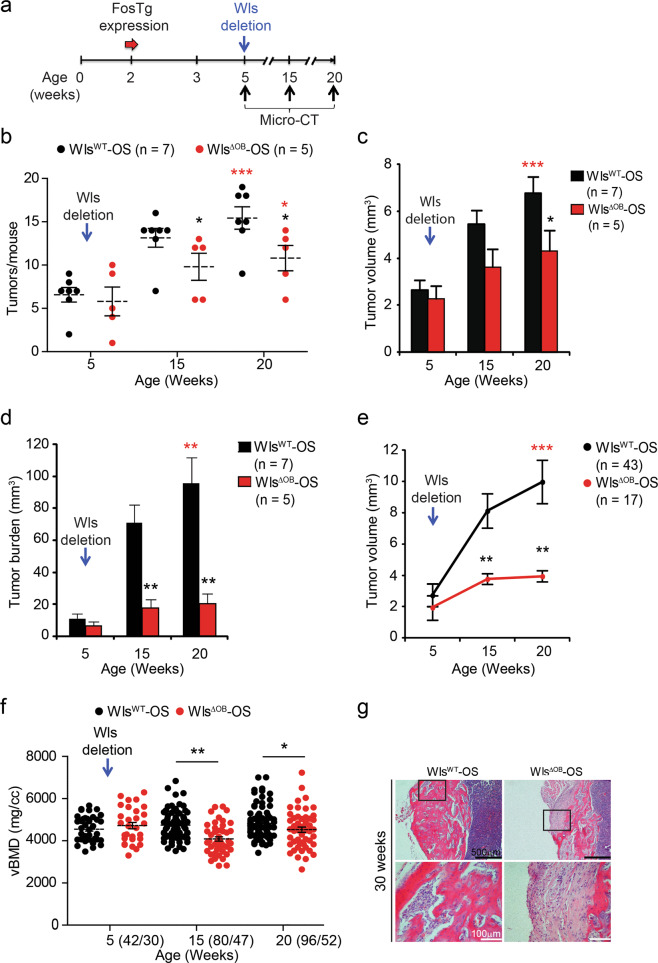
Fig. 3. Wls deletion during tumor progression decreases OS in H2-c-fosLTR mice.
a Experimental procedure. Dox is removed at 5 weeks of age when tumors are detectable by Micro-CT and the mice are additionally subjected to Micro-CT at 15 and 20 weeks. b–f Micro-CT analysis of WlsWT and WlsΔOB-OS mice. Tumor number per mouse (b), average tumor volume (c) and average tumor burden per mouse (d) at 5, 15 and 20 weeks in WlsWT-OS mice (n = 7) and WlsΔOB-OS mice (n = 5). e Volumetric follow-up over time for 43 WlsWT-OS tumors and 17 WlsΔOB-OS tumors. f vBMD in WlsWT-OS and WlsΔOB-OS tumors over time. The number of analyzed tumors is indicated in parentheses (WT/ΔOB) beside each time point. g Representative tumor histology at 30 weeks. Bar graphs and plots represent or include mean ± SEM, respectively. Black asterisks: *P < 0.05 and **P < 0.01 by two-way ANOVA with Bonferroni post hoc test. Red asterisks: ns not significant, *P < 0.05, **P < 0.01 and ***P < 0.001 by two-tailed t-test between each genotype at 20 weeks and all mice at 5 weeks.

