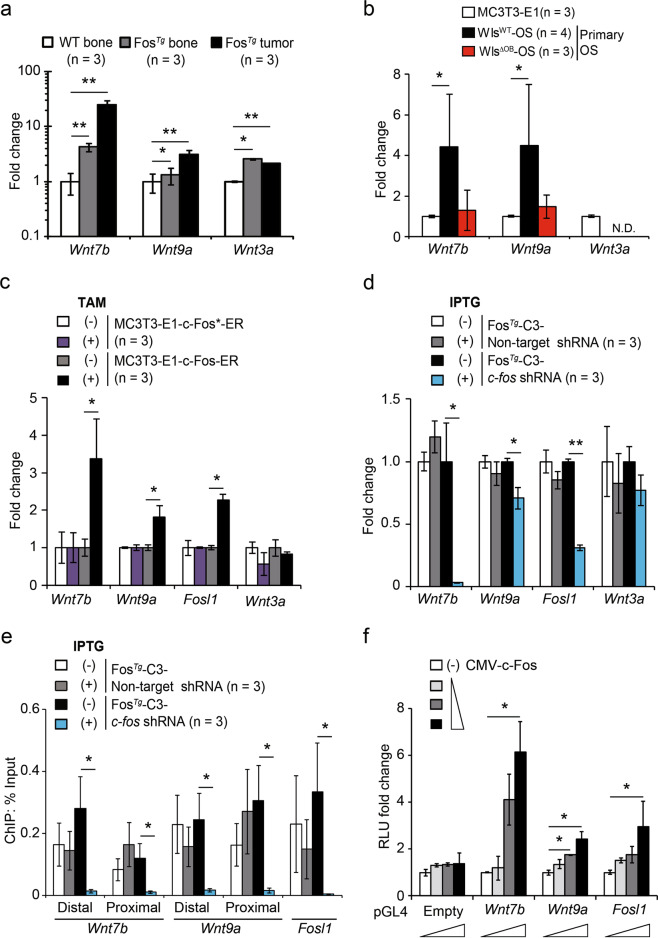
Fig. 4. c-Fos/AP-1 directly promotes the expression of Wnt7b and Wnt9a in OS.
a, b qPCR analysis of Wnt7b, Wnt9a and Wnt3a in H2-c-fosLTR bones, tumor-bearing bones and dissected tumors (a) and primary OS cells isolated from WlsWT-OS and WlsΔOB-OS mice (b). Bones from WT littermates (a) and MC3T3-E1 osteoblastic cells (b) were included for comparison. N.D., not detectable. c, d Gene expression in MC3T3-E1 cells expressing c-Fos-ER or mutant (inactive) c-Fos*-ER in the presence/absence of tamoxifen (c) and in the H2-c-fosLTR OS cell line (FosTg-C3) expressing IPTG-inducible c-fos shRNA or non-target shRNA in the presence/absence of IPTG (d) was determined by qPCR. FosL1 (encoding Fra-1 and a bona fide Fos-target gene) is included as a control. e ChIP-qPCR quantification of c-Fos/AP-1 binding to Wnt7b and Wnt9a promoters in H2-c-fosLTR OS cells expressing IPTG-inducible c-fos shRNA or non-target shRNA in the presence/absence of IPTG. The FosL1 genomic region with a c-Fos/AP-1 binding site is included as a control. f Luciferase reporter assay in MC3T3-E1 cells cotransfected with increased amounts of c-Fos expressing vector (CMV-c-Fos) and Wnt7b and Wnt9a reporter constructs containing the ChIP sites from e. A Fosl1 reporter construct is included as a control. Bar graphs represent mean ± SEM. *P < 0.05 and **P < 0.01.

