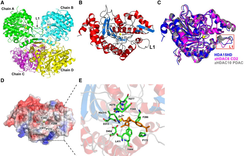
Figure 2.
Crystal structure of the HDA15HD tetramer and monomer. A, Ribbon diagram representation of the crystal structure of the HDA15HD tetramer in a substrate-free form. Zinc ions are represented by yellow spheres. L1, Loop structure (residues 161 to 174 of HDA15). B, HDA15HD monomer. The α-helices and β-sheets are colored in red and light blue, respectively. C, Superposition of HDA15HD (blue), zHDAC6 CD2 (magenta, PDB 5EEK), and zHDAC10 PDAC (gray, PDB 5TD7). The L1 loop is enclosed by a red frame. D, The active site of the HDA15HD domain is located at the center of the electrostatic surface with four conserved residues, P172, F286, Y344, and L411, around the cavity. E, The model of the HDA15HD–TSA complex showing the binding mode in the active sites with the conserved residues H276, H277, D313, H315, D404, and Y444. TSA is represented by brown.

